
This supporting evidence page contains the complete recorded AI responses used in Ask and AI Shall Find. The principal article presents selected excerpts for clarity. Expand any panel below to examine the full answer given for that image and prompt condition.
Reading note: These responses are reproduced as recorded. They are evidence from a small practical exercise, not verified biological identifications or professional authenticity determinations.
1. Blackwater larva
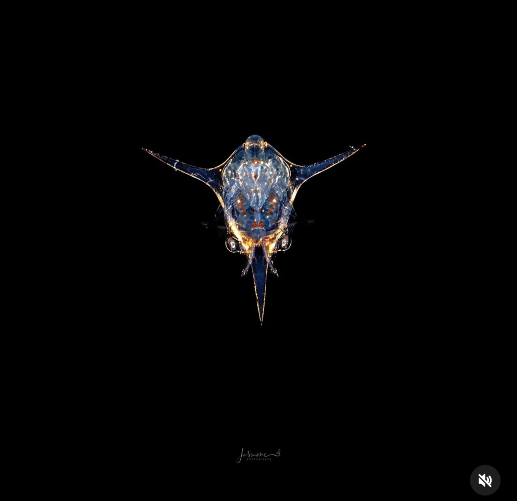
Prompt A – ChatGPT
No primary integrity red flag is visible, but this screenshot alone cannot authenticate the photograph.
Integrity verdict: Low visible suspicion, moderate confidence
1. Biological accuracy
This appears to be a crab zoea or closely related decapod larva photographed head-on. Its transparent
body, paired lateral spines and long carapace spine are biologically credible characteristics of zoeal
larvae. (Oxford Academic)
The animal is pelagic, so an open-water black background is entirely appropriate. There is no biologically
questionable host, substrate or behaviour.
2. Physical contact and support
There should be no footprint, contact shadow or substrate support. This animal naturally remains
suspended in the water column. Its apparent “floating” is therefore not suspicious.
The appendages and spines connect logically to the body. I see no detached structures, impossible
overlaps or inconsistent scale.
3. Optics and lighting
The strong highlights along the transparent carapace and spines are consistent with strobe light
refracting through a translucent subject. The lighting varies naturally across the body rather than
resembling a uniformly applied digital outline.
Focus and detail fall off slightly through the finer appendages, which is believable at this magnification. I
do not see an obvious extraction halo, repeated edge pattern or conflicting lighting direction.
The pure black background could conceal editing, but it is also a completely normal photographic result
during blackwater diving. Black alone is not evidence of compositing.
4. Manipulation assessment
There is no visible evidence that this is a digital composite, forced interaction or ecologically staged
photograph. However, this is a screenshot with the subject occupying a relatively small area. It is not
detailed enough to rule out edge cleaning, background repair, object removal or a captive setup.
For competition verification, I would examine the RAW file at 100 percent, the frames immediately before
and after it and the original capture metadata.
Photographic judgement
The frontal symmetry is excellent and the creature has immediate graphic impact. It reads like an alien
mask emerging from darkness. The pose is unusual enough to hold attention.
My criticism is that the subject is slightly too small within such an extensive black field. The negative
space is elegant, but there is more than the animal needs, particularly beneath it. A modestly tighter crop
would give the fine anatomy greater authority without losing the sense of open water.
Some lower central highlights are close to overpowering the transparent detail. If the RAW contains
recoverable information, slightly controlling those highlights would improve anatomical clarity.
Competition verdict: Credible and visually strong. No integrity-based rejection from the presented
image. A likely shortlist image for a Blackwater category, subject to RAW verification.
Prompt A – Claude
No primary red flag. This reads as a genuine blackwater-style capture of a planktonic crab zoea, with
one caveat: I can’t independently verify the pure black background from a compressed Instagram
screenshot.
Fake & Integrity Check
Biological accuracy: passes. The body plan fits a brachyuran (true crab) zoea larva: a glassy carapace, a
pair of long lateral spines, and a long central spine. The two rounded structures flanking the base of that
spine look like the compound eyes, so the animal is viewed dorsally with the rostrum pointing down. That
orientation is anatomically coherent. The red-orange spots are chromatophores, which are normal for
zoeae, and the faint out-of-focus appendages near the lower flanks are where thoracic limbs and
mouthparts should be. Zoeae are a few millimeters long and drift in open water, so a subject suspended in
nothing is ecologically correct, not a red flag. I can’t confirm the species from this angle. The lateral spines
suggest a portunid-type zoea, but that’s a hedge.
Physical contact and support: not applicable. A “floating” subject is what you’d expect here. There is no
substrate to cast a shadow on, and there should be none.
Optical and lighting consistency: mostly passes.
– Focus is crisp across the carapace and spine tips. The animal sits almost in one plane, so a thin macro depth of field is plausible. – The warm orange rim light along the lower edge and spine edges, plus cool blue transmission through the tissue, is consistent with strobe light from behind or below. Two light colors (a gelled or warm source plus a cool one) is possible, and I’d want the shooter to confirm it. – I see no halo, cutout fringe, or feathered edge around the spines or the translucent margins. Transparent edges are where composites usually fail, and these hold up. – The left and right sides are near-symmetric but not identical. The spine curves differ, and the small marks on each spine don’t mirror. That argues against a flipped or cloned half.
Manipulation assessment: no evidence of composite, with two caveats.
1. The background is featureless black, with no backscatter, marine snow, or gradient. That’s achievable with snooted strobes and a small aperture, but it’s also exactly what a masked background looks like. Compression hides the noise floor that would settle it. 2. Blackwater subjects are often gently restrained in a jar or container for a moment before shooting. That’s a normal ethical debate in this genre, not fakery, but I can’t tell from the frame whether it happened.
Photographic critique – Strengths: Real detail in the tissue and eye structures, a clean lighting split, and a strong subject with no distracting elements. – Weaknesses: The composition is a dead-center, symmetric placement with roughly 60% empty frame. It’s deliberate and graphic, but it reads static. The lower spine tip falls into shadow and loses definition, and the head area is slightly soft. The watermark is small and unobtrusive, which is fine.
What would settle it
The unedited original or RAW, the EXIF (aperture, shutter, strobe power), and the neighboring frames from
that dive. A real capture should show sensor noise and faint dust or particles in the “black” when levels are
lifted. A masked background usually won’t.
Prompt A – Gemini
The primary red flag in this image is its perfect bilateral symmetry, which strongly indicates a
digitally mirrored composite rather than a single, unedited raw capture of a planktonic organism.
Here is the integrity and technical audit:
– Biological Accuracy: The subject appears to be a decapod crustacean larva (likely a zoea larva of a crab, given the prominent dorsal and lateral spines). Planktonic organisms of this stage drift freely in the pelagic zone during blackwater dives, so suspended isolation against a pitch-black background is ecologically authentic for a water-column shot. However, true organisms are never 100% symmetrical down to internal organ structures, reflected glints, and fine setae. – Physical Contact & Support: As a pelagic plankter, the organism is neutrally buoyant in the water
column; a physical substrate or footprint is not required. However, there are no visible micro-particles, marine snow, or fluid motion artifacts surrounding the body, giving it a artificially sterile, laboratory-like isolation. – Optical & Lighting Consistency: The lighting (a mix of rim lighting and internal refraction/transillumination) is completely identical on both the left and right axes. The highlights on the lateral spines mirror each other with mathematical precision. The depth of field across the horizontal plane does not fall off naturally; both tips of the lateral spines retain identical sharpness and micro-refractions. – Manipulation Assessment: Digital Composite / Mirrored Image. This photograph has been cloned or mirrored across the vertical axis to achieve geometric symmetry. While symmetrical techniques are sometimes used in graphic design, in competitive underwater macro photography, unannounced axis-mirroring constitutes heavy digital manipulation.
Verdict: Stylized, aesthetically striking artwork, but fails strict authenticity standards for competitive
underwater photojournalism due to artificial bilateral mirroring.
Prompt B – ChatGPT
Overall assessment
The image appears biologically and photographically plausible. I see no clear visual evidence in this
screenshot that justifies calling it composited, staged, forced or AI-generated. However, a rendered
screenshot cannot establish authenticity.
1. Biological context
Direct observation: The subject is a small, transparent pelagic organism with a rounded carapace,
paired lateral spines, fine appendages and a long downward-pointing projection. It is suspended in open
water with no visible host or substrate.
Interpretation: The morphology is consistent with a crab zoea, an early planktonic larval stage. Crab
zoea are often translucent and spiny, although the precise species cannot be identified confidently from
this frontal view alone. (Shearwater Research Inc)
Nothing visible appears biologically impossible. Specialist examination and additional viewing angles
would be required for a reliable identification.
2. Physical contact and support
Direct observation: The animal has no visible contact with a substrate. There is therefore no contact
shadow or weight-bearing point.
Interpretation: This is normal for a planktonic larva suspended in the water column. Its apparent
“floating” is not inherently suspicious and does not require the kind of physical support expected from a
benthic animal.
The lateral structures are similar but not exact mirror images. Their angles, markings and highlights differ
slightly, which is compatible with a real three-dimensional subject viewed head-on.
3. Optical and lighting consistency
Direct observation:
– The central carapace and frontal anatomy hold the strongest detail. – Some finer appendages are softer and less distinct. – Bright highlights occur across the centre and along both lateral spines. – The transparent body contains irregular reflections, internal structures and colour. – The background is completely black with no visible bokeh or environmental detail. – I do not see an obvious cut-out boundary, repeated texture or uniform halo around the subject.
Interpretation: The lighting is consistent with close-range underwater strobes illuminating a transparent
subject against unlit open water. The softer appendages can reasonably be explained by shallow depth
of field, movement and their translucency.
Some highlights around the lower head appear very bright, but the screenshot does not contain enough
tonal information to determine whether they are clipped in the original. Likewise, the perfectly dark
background could result from ordinary blackwater exposure, deliberate black-point adjustment,
background cleaning or a combination of these.
4. Integrity questions
The following features merit routine verification, but are not evidence of wrongdoing:
– The exceptionally clean black background could conceal removed particles or backscatter. – The strong symmetry may initially appear unusual, but a head-on view provides a straightforward photographic explanation. – The transparency and bright edge reflections make accurate masking difficult, yet I see no clear masking artefact at this resolution. – The file supplied is a screenshot with a watermark and interface icon, not the original image. Compression and resizing could both conceal genuine detail and create misleading edge effects.
Provisional conclusion: I see no specific visual contradiction suggesting compositing, AI generation or
forced staging. The image should not be labelled manipulated on the evidence available.
Aesthetic feedback
The frontal perspective is striking. The lateral spines create a strong horizontal shape while the long
rostral spine gives the composition a vertical anchor. Warm internal highlights against the cool
transparency make the animal feel intricate and almost mechanical.
The extensive negative space supports the isolated, open-water character, although a slightly tighter
presentation could give the subject more immediate impact. The darkest internal areas and brightest
highlights should be checked in the original file to ensure delicate anatomical detail has not been lost.
Evidence needed for further investigation
– The original RAW file – A full-resolution final export rather than a screenshot – EXIF and capture metadata – Adjacent frames showing changes in position and appendage movement – The unedited frame for comparison with the final image – Lightroom, Capture One or Photoshop editing history, including any layers – Disclosure of backscatter removal, background darkening or object removal – The photographer’s account of the dive, subject encounter and lighting arrangement – Field observations from a guide or other diver, if relevant – Identification by a specialist in decapod larval morphology, supported by additional viewing angles
Prompt B – Claude
Review: transparent planktonic organism on a black background
This is a screenshot of a social media post. The mute icon at bottom right suggests it may be a video or Reel
frame. That limits what I can say about noise, halos and fine edges, because platform compression and the
screenshot process both degrade exactly the detail an integrity check depends on.
1. Biological context
Observed: A single, mostly transparent animal is centred in the frame with no visible substrate, host or
other organism. It has a roughly oval body with internal structure, two long lateral spines, one long tapering
spike at the bottom, and a small dome at the top. There are orange-red pigment spots inside the body,
likely chromatophores, and paired rounded structures near the base that may be eyes. Very faint, thin
structures extend sideways at about mid-body height on both sides.
Interpretation: The combination of a transparent body, red chromatophores and multiple long spines is
consistent with a decapod crustacean larva, possibly a brachyuran (crab) zoea. Swimming-crab zoeae are
the kind that often show a rostral spine plus lateral spines. This is a plausible fit, not an identification. The
viewing angle is ambiguous, so I can’t say confidently which spike is rostral or dorsal, or which end is
anterior. Other larval forms can look superficially similar.
Association plausibility: There is no host or substrate, so the question doesn’t apply. A free-swimming
larva suspended in open water is biologically normal. This looks like a pelagic or “blackwater” style subject.
Confirming the taxon needs a larval-crustacean specialist, ideally with more angles.
2. Physical contact and support
Observed: There are no visible contact points, shadows, tethers, or surfaces. Nothing appears to be
holding the animal.
Inferred: The animal is presumably suspended in the water column, which fits its biology. From one
two-dimensional frame I can’t rule out an unseen fine support, such as a jar, a hand-held specimen
container, or a cropped-out edge. Hand-placed or contained specimens exist in photography, and this
image can’t confirm or exclude them. Scale is also unknown. Zoeae are usually a few millimetres, but
nothing in the frame confirms that.
3. Optical and lighting consistency
Observed:
– Focus is fairly uniform from the tip of the lateral spines to the central body. The lower spike loses a little definition toward its tip. – The lighting is split by colour: cool blue-grey across the body and spines, with strong amber-orange rim light along the lower body edge, on the upper edges of the lateral spines, and at the tip of the right spine. – The background is a uniform black with essentially no backscatter or drifting particles. I can see only one extremely faint speck below the animal, at about the 530, 870 pixel position. – The edges look clean against the black, and I see no obvious halo at this resolution.
Ordinary explanations:
– Uniform focus is normal for a small subject that lies roughly in one plane, shot at a small aperture, or focus-stacked. A transparent, planar animal is easy to keep in focus. – The warm and cool split is plausible from a transparent body refracting and dispersing light, or from a backlit or rim-lit setup with a narrow light source or snoot. – A very clean black is common in blackwater and snooted-strobe work, where the beam illuminates only the subject. Heavy black-point adjustment in editing has the same effect.
Possible-manipulation explanations: Strong background darkening, masking, or colour grading could
produce the same look. I can’t distinguish these possibilities from this image.
4. Integrity questions to investigate
These are questions, not conclusions.
1. Near-total absence of particles. Night pelagic dives usually show some marine snow or backscatter. Its absence could come from narrow lighting or from black-point clipping or masking. 2. Strong bilateral symmetry. Symmetry is expected in a larva seen head-on or dorsally, so it isn’t suspicious in itself. However, small asymmetries argue against a simple mirrored duplicate. The two lateral spines differ in tip detail, and there seems to be a small bead-like feature on the left spine with no counterpart on the right. The faint side appendages also differ. It would still be worth checking the finest structures at full resolution for repeated patterns. 3. Warm and cool colour split. Check whether the amber comes from the light source or refraction or was introduced by split-toning or selective grading. 4. Centred subject on a large black field. This may be a crop or a placement on a black canvas for social media. That is common presentation practice and not manipulation on its own, but it’s worth confirming whether the subject was extracted or the frame is original. 5. Faint side structures. Check whether they are real out-of-focus or dimly lit appendages, or remnants of a mask edge.
I see nothing here that supports calling the image composited, staged, or AI-generated. The detail looks
anatomically coherent and internally consistent, and the flaws that would indicate those things (warped
anatomy, inconsistent spine geometry, smeared internal structure) aren’t apparent. That is a limited
statement given the image quality.
Aesthetic notes
The composition is strong. Centred symmetry, generous negative space and the blue-and-amber contrast
give it a jewel-like quality. The main technical critique is that the subject is small in the frame, so if the
original resolution allows a tighter crop, more of the internal detail would be visible. The watermark is
discreet and well placed.
Evidence that would help a human reviewer – The original RAW file and full-resolution export, to inspect noise, edge behaviour and clipping. – EXIF data (aperture, shutter, ISO, strobe settings, lens and any diopter or wet lens). – Adjacent frames from the same encounter. If this is a video frame, the source video would be ideal, since it would show the animal’s motion and rotation. – The editing history or an unedited JPEG straight from the RAW conversion. – Depth, location and dive type (blackwater, night, day), and whether the specimen was free-swimming or contained. – Field notes or the photographer’s account of the encounter. – Review by a specialist in larval crustaceans for identification.
Prompt B – Gemini
Biological Context – Visible Organism: The image depicts a highly translucent marine planktonic organism set against a pure black background. The bilateral symmetry, prominent lateral spine-like extensions, pointed posterior projection, and transparent carapace strongly resemble a larval stage of a decapod crustacean (such as a zoea or megalopa stage of a crab) or a specialized pelagic mollusk (like a cavolinid pteropod/sea butterfly). – – Host/Substrate Association: The subject is floating freely in open ocean water (blackwater context) with no substrate, host, or physical anchor visible in the frame. – – Plausibility: Planktonic organisms of this morphology are commonly encountered during night dives in deep water (blackwater photography). The bilateral geometry and internal organ structure visible through the transparent shell are biologically consistent with larval marine plankton. –
Physical Contact & Support – Observable Points of Contact: There are no visible physical contact points, resting surfaces, or tethering structures supporting the organism. – – Shadows & Weight Distribution: No cast shadows exist due to the total absence of a adjacent background surface. The organism appears entirely suspended in mid-water. – – Observation vs. Inference: Direct observation shows a centered, symmetrical, floating structure surrounded by uniform pitch black. From this two-dimensional frame alone, it cannot be determined whether the organism was freely swimming in place, drifting passively in a current, held by a microscopic tether removed in editing, or isolated digitally via a background brush/mask. –
Optical & Lighting Consistency – Focus Plane & Depth of Field: The focal plane is exceptionally narrow, passing cleanly through the central core and lateral spines. The anterior/posterior edges show subtle fall-off into soft focus consistent with high-magnification macro lenses (such as a 60mm or 105mm macro combined with a wet diopter). – – Lighting Direction: High-contrast rim lighting and internal refraction are evident. Warm, orange-gold
backlighting/side-lighting illuminates the lower interior organs and lower margins, while cool blue rim lighting highlights the outer lateral spines. – – Edge Transitions & Bokeh: The perimeter edges of the organism transition directly to pure RGB black (0, 0, 0) without visible backscatter or ambient water column noise. The lack of any out-of-focus suspended particles (marine snow) around the subject is unusual for a raw underwater shot, though achievable with ultra-narrow snoot lighting, strong negative exposure, or heavy global black-point crushing in post-processing. – – Optical Explanations vs. Manipulation: The high contrast and total isolation could be the result of a tight optic snoot/strobe setup designed to illuminate only the subject. Alternatively, it could indicate background removal or aggressive masking during post-processing to clean up marine snow. –
Integrity & Investigative Features – Features Warrants Investigation: 1. Uniform Pure Black Background: The total uniform blackness devoid of backscatter or water texture. 2. Bilateral Symmetry Alignment: The image features nearly flawless central alignment, which can occur naturally in live subjects, but can also be the result of vertical mirroring or geometric cropping/alignment adjustments. 3. High-Contrast Internal Glow: The intense internal orange/gold refraction should be evaluated to verify if it stems from targeted fiber-optic strobe placement or selective saturation/luminescence local adjustments. – Proportionate Finding: There is no conclusive visual proof of digital compositing, AI generation, or physical staging based on this single flattened JPG file alone. The features observed fall within the boundary of advanced blackwater macro technique, heavy post-processing cleanup, or a combination of both.
Evidence Required for Further Investigation
To definitively verify the authenticity, capture method, and editing scope of this photograph, a reviewer would
require:
1. Unedited RAW File (.CR3, .NEF, .ARW, or .DNG): To evaluate sensor-level light capture, true optical fall-off, and raw sensor dynamic range before post-processing curves were applied. 2. Complete EXIF / MakerNote Metadata: To confirm exposure settings (aperture, shutter speed, ISO), lens focal length, strobe fire status, camera model, and original file creation timestamps. 3. Adjacent Burst/Sequence Frames: Original sequential shots taken before and after this exposure to observe the subject’s natural movement, drift, and orientation in the water column. 4. Non-Destructive Project File (PSD/TIFF with Layers or Lightroom Sidecar): To examine the exact editing history, such as the use of subject masks, background cleanup brushes, localized saturation adjustments, or cloning tools. 5. Photographer’s Field Account: Details regarding the dive site (location, depth, blackwater drift), lighting gear (e.g., fiber-optic snoots, backlights), and technique used during capture.
2. Bearded goby
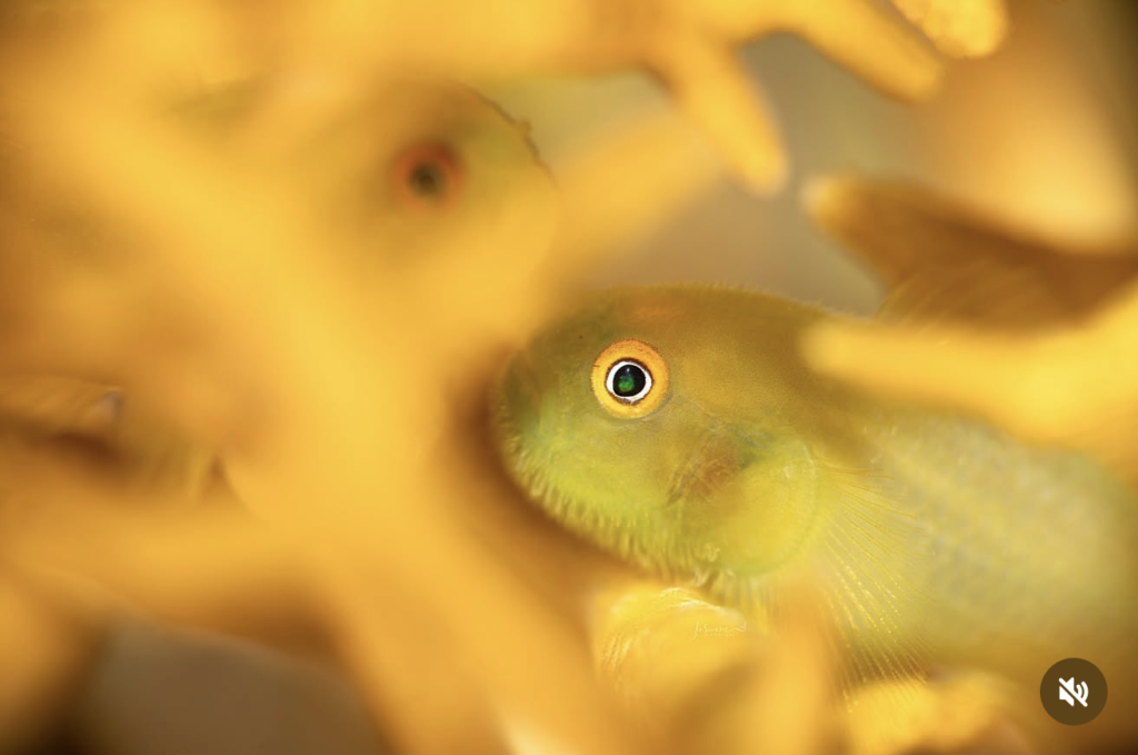
Prompt A – ChatGPT
No primary integrity red flag is apparent. The image is biologically and optically coherent, although the
exact goby and host cannot be confirmed from the photograph alone.
Biological accuracy: The small goby is positioned naturally within a branching coral or similar reef
organism. Many gobies live closely among coral structures, so the association is plausible.
(DivePhotoGuide) The second fish behind it is also believable, suggesting a pair sharing the same
shelter.
Physical contact: The main fish appears supported between the surrounding branches, with its lower
body and fin resting naturally. It does not look suspended or pasted into the scene.
Optics and lighting: The primary eye occupies the sharpest focus plane while the face, body and
second fish become progressively softer with distance. That depth transition is logical. Lighting direction
and warm colour remain consistent across both fish and the host. The extremely crisp eye may have
received selective sharpening, but it is not evidence of compositing.
Manipulation assessment: No visible extraction halo, conflicting edge, duplicated feature or lighting
mismatch suggests a composite. Colour saturation and warmth appear heavily developed, but that is
tonal processing rather than structural manipulation.
Photographic judgement: The eye is magnetic and the monochromatic yellow palette is distinctive. The
blurred second eye adds a quiet narrative. However, the intense yellow processing suppresses texture
and makes the foreground feel slightly heavy. The main fish’s body also exits the frame rather abruptly
on the right.
Verdict: Visually credible and competition-worthy. Stronger artistically than technically perfect, with
colour restraint and slightly more breathing room on the right likely improving it.
Prompt A – Claude
No primary red flag. This looks like a genuine shot of a coral goby (Gobiodon) shot through
out-of-focus coral branches, but I can’t tell from a compressed screenshot whether it was taken on
a reef or in an aquarium.
Fake & Integrity Check
Biological accuracy: passes.
– Coral gobies are obligate branching-coral dwellers, so a small goby wedged between coral branches is the expected habitat. The fish’s yellow-green coloring against yellow-orange coral is also consistent with the coral-matching color morphs in this genus. – The blurred second fish in the upper left, showing only an orange-red eye, fits too. Gobiodon live in pairs or small groups within a single colony. – The size relationship works: the fish is only a few centimeters long and sits comfortably among the branches. – I can’t name the species from this angle. The eye ring and green body point to a green or yellow Gobiodon morph, and I’d want the head-line markings visible before committing. The coral looks like a digitate Acropora or similar, but it’s too blurred to confirm.
Physical contact and support: passes. The fish is nestled inside the branch structure with coral in front of
and behind it, so there’s no floating problem. The pectoral fin rays on the right fan out against the coral and
dissolve into blur at a believable rate.
Optical and lighting consistency: mostly passes, with two things I’d check.
– The focal plane sits on the eye and cheek, and everything nearer the lens dissolves into smooth bokeh. Shooting through blurred foreground elements also produces a milky veil over the subject, and the fish’s right side and fin do show that soft, low-contrast wash. That is what genuine foreground blur does, and it’s hard to fake convincingly. – The lighting is warm and coherent across the frame, with no conflicting shadow direction. – Check 1: The fine “fuzz” along the cheek and chin outline is unusually dense. Gobies do have sensory papillae, so this could be real. At this resolution it could equally be oversharpening, a clarity or texture slider, or noise reduction. I’d want a 100% crop to tell. – Check 2: There’s a soft luminous haze along the top edge of the head. It could be the foreground veil, but it’s also where a masking halo would show up. I see no hard cutout edge, so I lean toward natural.
Manipulation assessment: no evidence of composite. Two caveats.
1. Wild or captive? The smooth, neutral beige-grey background has no blue water and no reef context. Strobe-lit sand or rock could produce that, but so could a tank wall. Coral gobies are among the most photographed aquarium fish, and this frame is consistent with either setting. If it was shot in a tank, that’s legitimate work, but it should be captioned that way. 2. Color grading: The image is almost monochromatic yellow-orange, so a heavy white-balance or saturation push is likely. That’s a stylistic choice, not fakery, but it makes the color relationships harder to trust.
Photographic critique – Strengths: The eye is razor sharp with clear catchlights and a rich green pupil reflection. The blurred foreground branches create a “peeking through” frame that adds depth. The second out-of-focus fish adds narrative, and the color palette is cohesive. – Weaknesses: The foreground yellows are close to blown, with little tonal separation, so the branches read as flat blobs. The bright blob at bottom center pulls the eye away from the subject. The eye sits almost dead center, which weakens the layered composition. The fish’s body behind the
right-hand branch turns pale and muddy, losing form. The dense edge fuzz needs to be explained or cleaned up.
What would settle it
A full-resolution crop of the head and cheek would show whether the fuzz is papillae or processing. The
EXIF (aperture, strobe setup) and the neighboring frames would show whether the background is a tank
wall or reef. If it’s a tank, the caption should say so.
Prompt A – Gemini
The primary red flag in this image is the presence of a “ghost” eye in the out-of-focus yellow
coral branch to the left, which strongly suggests either an artificial multi-exposure, a digital cloning artifact,
or a composited overlay of another fish.
Here is the integrity and technical audit:
– Biological Accuracy: The subject appears to be a yellow coral goby (Gobiodon okinawae or similar species) nestled inside the branches of an Acropora or soft/leather coral host. This is an ecologically accurate interaction, as these gobies obligately inhabit live coral colonies, using the branches for protection, breeding, and perching. – Physical Contact & Support: The goby’s pectoral fin and ventral body are naturally resting against the lower coral stalk, and its body conforms logically to the curvature of the host. The physical footprint looks authentic to how these small gobies wedge themselves inside coral structures. – Optical & Lighting Consistency: The main subject shows sharp focal plane detail on the eye, pectoral fin rays, and skin texture, transitioning into a very shallow depth of field. The warm ambient lighting on the fish matches the golden hue of the foreground and background coral. However, the blurred region directly to the left contains a distinct dark ring with an orange center that mirrors the color and geometry of the primary goby’s eye, creating an optically unnatural “double eye” effect that does not align with standard lens bokeh or depth-of-field blur. – Manipulation Assessment: Highly Suspicious / Potential Digital Edit. While the goby itself is physically real and properly situated in its habitat, the secondary eye shape in the left-hand blur is a major anomaly. This is likely a result of: 1. A double-exposure in-camera where a second goby (or the same goby) shifted position. 2. A digital composite/clone tool error when trying to remove unwanted debris or smooth out the foreground bokeh. 3. A second goby deep in the background whose body is entirely obscured, though its scale and position relative to the blur render this visually improbable.
Verdict: Environmentally and biologically sound in concept, but compromised by a glaring optical/digital
artifact in the left foreground that invalidates it as a clean, unedited single frame.
Prompt B – ChatGPT
Biological context
Observation: A small yellow-green goby is positioned among branching coral, with a second blurred fish
visible behind it.
Interpretation: The subject is consistent with a coral goby, likely Gobiodon or a related genus. These
fishes commonly live within branching corals, so the association is biologically plausible. The precise
species and coral cannot be confirmed from this image alone.
Physical contact and support
Observation: The foreground fish appears nestled between coral branches. Its lower body and fins are
partly obscured, so the exact contact points are not visible.
Interpretation: Its position and body angle are consistent with a small goby resting within coral. Nothing
suggests unnatural suspension. The hidden contact points are readily explained by the viewpoint and
shallow depth of field.
Optical and lighting consistency
Observation: Focus is strongest on the foreground eye and nearby facial detail. Sharpness falls
progressively across the body, coral and second fish. The foreground edges are soft but continuous,
without an obvious cut-out halo. The green eye reflection is contained within the pupil.
Interpretation: The focus transition is consistent with close-range macro photography at a wide
aperture. The eye reflection is plausibly produced by strobe or focus-light reflection. Warm illumination is
reasonably consistent across the fish and coral.
Integrity questions
The second fish should be checked in adjacent frames to confirm its changing position, although it does
not appear to be a simple duplicate. The strongly softened coral could conceal local editing, but there is
no specific visual evidence of compositing or AI generation.
Aesthetic assessment
The sharply rendered eye provides an immediate focal point, while the second fish adds depth and
context. The nearly monochromatic yellow palette feels cohesive. The foreground coral is quite dominant
and obscures much of the fish, but it also creates an intimate, habitat-based perspective.
Helpful evidence: RAW file, full-resolution export, metadata, adjacent frames, editing history and the
photographer’s account of the encounter.
Prompt B – Claude
Review: coral-dwelling goby framed through out-of-focus branches
As with the last image, this is a screenshot with a mute icon, so it is probably a frame from a Reel or video.
Platform compression, and possibly zoom or pan effects, limit what I can say about fine edges and noise.
1. Biological context
Observed: A small yellow-green fish sits in the focal plane, with a sharply rendered eye. The eye has an
orange outer ring, a pale inner ring, and a dark pupil with a green glint. The head and cheek carry fine
bristle-like or papillate texture, and pectoral fin rays are visible on the right. The fish is surrounded by
heavily blurred, blunt-tipped, finger-like branches in saturated yellow-orange, which look like branching
coral. A second, fully blurred fish head with an orange-red eye sits in the upper left. Behind them is a pale
grey-beige background.
Interpretation: The body shape, small size, yellow-green colouring and position tucked between branches
are consistent with a coral-dwelling goby, probably Gobiodon or possibly Paragobiodon. These fish live in
branching corals such as Acropora, Pocillopora and Seriatopora, often match the colour of the host, and
are frequently found in pairs. The second blurred fish therefore fits the biology. I can’t identify the species.
The pattern and head details that separate the candidates aren’t resolvable here, and the coral genus can’t
be determined from the blur.
Plausibility: The association is plausible. A yellow-green goby within yellow branching coral is a well-known
pairing. This is an interpretation, and a fish specialist would need to confirm it.
2. Physical contact and support
Observed:
– Foreground branches overlap the fish’s body on the right and the snout on the left, so the fish appears to sit behind and between branches. – A blurred branch stub lies just below the chin. – I see no tweezers, hand, tether, container edge or other support. – Shadows are soft and weakly defined, so the lighting gives few cues about depth or contact.
Inferred: The fish is probably resting or perching within the colony, as these gobies do. I can’t see the pelvic
fin or sucker disc, so I can’t say where it is actually anchored. From one 2D frame I also can’t measure how
far the foreground branches sit in front of it. Scale isn’t confirmed, though fish of this type are usually a few
centimetres long.
3. Optical and lighting consistency
Observed:
– Depth of field is extremely shallow. Focus sits on the eye and adjacent cheek, then falls off toward the snout and along the body to the right. – The foreground blur is smooth and translucent, and it veils parts of the sharp fish rather than cutting hard edges across it. – The overall light is warm, soft and fairly directionless, with no hard shadows or strong specular highlights. – The image has a diffuse, hazy glow. Contrast falls off toward the right side of the body. – The background is pale grey-beige, not the blue or green typical of open reef water.
Ordinary explanations:
– Semi-transparent foreground blur is what you get shooting wide open through a nearby branch that is narrower than the effective aperture. Composites more often show sharp mask edges or uniformly opaque blur. – The focus falloff is consistent with a fish angled to the lens plane at high magnification. – A pale background can come from a close sandy or dead-coral surface lit by strobe, or from white balance adjusted to neutralise the water colour. – The warm haze could come from a diffused strobe, veiling glare or lens flare, or from post-processing such as dehaze, negative clarity or a glow effect.
Possible-manipulation explanations: Selective glow or softening, heavy warm grading and local
sharpening could produce parts of this look. I can’t separate those from optical effects using this frame
alone.
4. Integrity questions to investigate
These are questions, not conclusions.
1. Bristly fringe around the head. Fine radiating spikes along the silhouette are sharp and dense. They may be genuine sensory papillae or mucus, but they could also be sharpening or texture-slider artefacts. Comparing this with edge behaviour on the fin rays and the eye at full resolution would help. 2. Haze and low contrast on the right body. It may be natural veiling from the foreground or an added glow. Check whether the falloff follows the optical geometry.
3. Wild or captive setting. The pale, evenly lit background and warm cast leave open whether this is a reef, a tank or a controlled setup. Nothing visible confirms either, such as glass reflections or tank edges. This matters for how the image is presented as wildlife photography. 4. Colour grading. The image is strongly warm and yellow-dominant. Check whether the yellow channel is clipped, and whether the fish’s yellow-green versus the coral’s orange-yellow is a natural difference or a grading choice. 5. The second blurred fish. It looks natural, and there’s no sign of a duplicate. It is too blurred for me to assess beyond that, and the different eye appearance is what blur would normally produce. 6. An indistinct dark structure at left-centre. I can’t interpret it. It may be a fin, shadow or another organism.
I see nothing that supports calling this composited, staged or AI-generated. The eye structure, fin rays and
overlap of foreground and subject look coherent, and the blur behaves like real optical blur. That statement
is limited by the compression and the soft, glowing rendering.
Aesthetic notes
The image is strong. The blurred branches act as a natural frame and vignette, the sharp eye gives an
immediate focal point, and the second blurred fish adds a sense of pairing. The near-monochrome warm
palette is cohesive, and the tiny green pupil glint is a good accent.
The main critiques are that the haze softens the fish’s body and reduces contrast where it meets the
foreground, and that strong yellow saturation risks losing tonal separation. A slightly more open
composition on the left might also relieve the crowding from the blur.
Evidence that would help a human reviewer – The RAW file and unedited JPEG, to inspect the haze, fringe texture and channel clipping. – EXIF data: aperture, focal length, magnification, strobe or diffuser setup, and white balance. – Adjacent frames or the source video, especially any showing the fish’s movement or the coral before and after framing. – The editing history, including dehaze, clarity, glow, sharpening and selective adjustments. – Location, depth and habitat, and whether it was shot in the wild or in an aquarium. – The photographer’s account of how the shot was made, including any coral moved or repositioned. – Identification of the fish by a goby specialist and of the coral by a coral taxonomist.
Prompt B – Gemini
Biological Context – Visible Organisms & Substrate: The primary focal subject is a small, greenish-yellow fish featuring a distinctive patterned iris and opercular details, nestled within the branches of a stony hard coral (likely a species of Acropora or Seriatopora). A second, out-of-focus subject appears in the upper-left quadrant, showing a similar dark eye/marking through blurred coral branches. – Plausibility: The primary subject strongly resembles a coral goby (genus Gobiodon, such as Gobiodon histrio or Gobiodon citrinus). These small gobies are obligate coral dwellers that spend their lives sheltered deep inside branching hard coral colonies. The biological association depicted between this goby species and the branching coral host is highly authentic and ecologically accurate.
Physical Contact & Support – Observable Points of Contact: The lower jaw, ventral area, and pectoral fin of the primary goby appear in direct physical resting contact with the yellow coral polyps/branches. – Shadows & Weight Distribution: Light casting beneath the fish’s belly and lower jaw creates natural, soft ambient shading onto the coral tissue directly beneath it. The fish’s body contours yield naturally to the coral structure, consistent with a small organism resting on a solid surface. – Observation vs. Inference: Direct observation shows the fish nestled within foreground and background coral branches. From this single frame, it can be inferred that the fish is comfortably wedged inside its home colony; whether it was actively moving or stationary at the moment of capture cannot be confirmed without video or burst frames.
Optical & Lighting Consistency – Focus Plane & Depth of Field: The focal plane is razor-thin and accurately placed right on the cornea and iris of the primary goby’s eye. The snout, cheek, and pectoral fin show progressive, natural optical fall-off. – Foreground Blur & Bokeh: Extreme macro foreground bokeh is present. The smooth yellow shapes wrapping around the frame represent coral branches positioned extremely close to the front element of the lens (shooting “through” the reef structure), creating a classic framing technique. – Lighting Direction: Diffuse, warm lighting illuminates the scene uniformly from the front/upper-right. Catchlights in the eye align logically with ambient underwater strobe flash or video lights. Edge transitions between the sharp eye/skin of the fish and the soft foreground coral branches exhibit natural optical roll-off with no hard halos or jagged masking artifacts.
Integrity & Investigative Features – Features Warrants Investigation: 1. Upper-Left Element: The blurred, circular dark mark in the upper-left quadrant. While likely a second goby deeper in the coral head or an out-of-focus polyp structure, its heavy blur warrants checking adjacent frames to confirm its identity. 2. Color Saturation: The rich, uniform golden-yellow tones throughout the frame should be evaluated to determine if global color temperature adjustments or selective saturation boosts were applied in post-processing to enhance the high-key visual effect. – Proportionate Finding: The visual evidence strongly aligns with authentic optical physics-specifically high-magnification macro photography using wide apertures (/2. 8 to /5. 6) shot through tight structural openings. There are no clear visual indicators of digital compositing, AI synthesis, or unnatural physical displacement.
Evidence Required for Further Investigation
To fully verify the technical capture and editing scope of this macro photograph, a reviewer would look for:
1. Unedited RAW File (.CR3, .NEF, .ARW, or .DNG): To verify original white balance settings, true optical depth-of-field, and uncompressed edge detail around the foreground coral branches. 2. Complete EXIF Metadata: To review aperture value, focal length, lens model, shutter speed, ISO, and strobe firing mode. 3. Burst/Sequence Shots: Consecutive frames to confirm the movement of the goby within the coral head and verify the second out-of-focus element in the background. 4. Post-Processing Layer File (PSD/TIFF or XMP sidecar): To check if local brush adjustments, Dodge/Burn tools, or color grading passes were used to clean or enhance the high-key yellow tones. 5. Photographer Context: Confirmation of whether artificial lighting (e.g., dual strobes with diffusers or continuous focus lights) was used to achieve the soft illumination within the dense coral head.
3. Creative seahorse
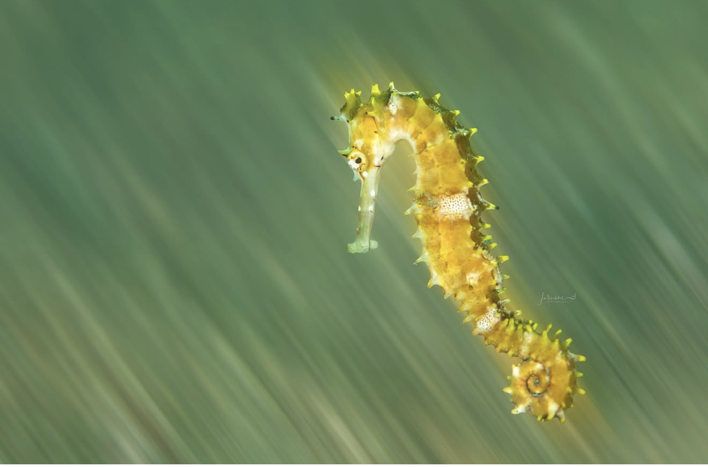
Prompt A – ChatGPT
Primary red flag: the seahorse has no visible holdfast and appears suspended in open water, so the
photograph cannot establish whether it was swimming naturally or had been dislodged.
Biological accuracy: The anatomy, colour variation and curled prehensile tail are biologically plausible.
Seahorses can swim between perches, so this position is possible. However, they commonly remain
anchored and are weak swimmers. The absence of a holdfast deserves scrutiny, particularly in an
ethics-led competition.
Physical contact: There is no substrate contact, weight-bearing point or attachment. That is consistent
with a brief swimming moment, but the image alone cannot distinguish natural movement from a staged
release.
Optics and lighting: The sharply illuminated animal against a directionally streaked background initially
looks suspicious. However, the consistent diagonal movement and faint trails around several body
projections support a credible slow-shutter, camera-movement and strobe-freezing technique. The
separation does not automatically indicate masking. Some bright areas on the head and body approach
lost highlight detail.
Manipulation assessment: I see no decisive extraction halo or mismatched lighting proving a
composite. Digital background blur remains possible, but the visual evidence also supports an in-camera
technique. The RAW file and adjacent frames would resolve this quickly.
Photographic judgement: Strong directional movement, effective subject placement and excellent
negative space in front of the face. The technique turns a conventional seahorse portrait into something
energetic. The green-yellow palette is slightly heavy and some fine anatomical detail is sacrificed.
Verdict: Visually strong and technically plausible, but ethically unverified. I would not reject it from the
finished image, although I would request the RAW sequence and capture explanation before advancing
it in a competition.
Prompt A – Claude
Primary red flag: a razor-sharp subject on a perfectly uniform linear-streak background is either a
legitimate strobe-plus-slow-shutter camera-movement shot or a digital motion-blur composite, and
I can’t confirm which from this screenshot. The tail is also curled around nothing.
Fake & Integrity Check
Biological accuracy: passes, with one oddity.
– The body plan is a spiny seahorse, most likely a thorny seahorse (Hippocampus histrix), given the long, pointed body spines, the coronet, and the white spotted patches on the trunk. I can’t rule out a close relative from a single side view, so treat that ID as a hedge. – The size and posture are plausible, with a curved neck, a downward-angled snout, and a small eye. – The oddity: the tail is coiled tightly but grips nothing. Seahorses are weak swimmers and spend most of their time anchored to seagrass, coral, or sponge. A free-floating adult happens when one is drifting, dislodged, or handled. Here the coil looks like it was gripping something that isn’t there. That leaves three possibilities: a genuine drift, a seahorse moved off its holdfast for the shot, or a holdfast removed in editing. I can’t distinguish them from this frame.
Physical contact and support: flagged. The animal has no visible contact point, no shadow, and nothing
to cast one on. In open water that’s acceptable, but combined with the empty tail coil it’s the weakest point
of the image. It needs an explanation.
Optical and lighting consistency: ambiguous, and this is the crux.
– The subject is crisp down to the individual spine tips, with no directional smear on its own edges. The background is dragged into long diagonal streaks at one consistent angle. – Consistent with a real capture: a slow shutter while moving the camera, with the strobe firing to freeze the subject, produces exactly this. The warm yellow glow along the seahorse’s right side could be the faint ambient-light ghost of the subject smeared across the frame during the exposure, which is what this technique leaves behind. – Consistent with a digital filter: the streaks are strikingly uniform in width, length, and angle from corner to corner. Camera movement through a real three-dimensional scene usually shows some variation with distance, and rotation or zoom blur would fan out radially. This looks close to a flat directional motion-blur filter. The glow around the body could also be a mask halo, an Orton-style glow, or a dodge layer. – I don’t see a hard cutout edge or feathering around the spines. The spiky silhouette would be very
hard to mask cleanly, and the spines look intact, which counts in the photographer’s favor.
Manipulation assessment: unresolved, leaning technique-based. The image is at minimum a stylized,
technique-driven shot. Whether the streaks are in-camera or applied later is a disclosure question in most
competitions. If the background was added or filtered digitally, this is a composite or heavy edit and should
be labeled that way. The empty tail is the second thing I’d ask about.
Photographic critique – Strengths: The diagonal composition with the subject tilted off-center gives real energy. The yellow-orange subject stands out cleanly against the muted green, and the spine detail and textured spots are excellent. The spiral tail makes a strong secondary focal point. – Weaknesses: The snout looks pale and near-blown, and the warm glow behind the body reduces edge contrast. The uniform streaking is arguably too clean, which is exactly what makes it look filtered. The watermark sits over the streaks and competes with the pattern.
What would settle it – The RAW file, or the neighboring frames from the same sequence. An in-camera pan shows frame-to-frame variation in streak shape, and a filter gives identical streaks each time. – The EXIF: a shutter speed around 1/4 to 1/2 s with a strobe would support the in-camera claim. A fast shutter with these streaks would be a red flag. – A 100% crop of the body edge, to see whether the glow is a directional ghost trail or a symmetric halo. – The photographer’s account of what the seahorse was gripping and how it ended up there.
Prompt A – Gemini
The primary red flag in this image is a obvious light-colored halo and edge cutout artifact
outlining the entire seahorse, which unequivocally confirms a heavily masked digital composite or an
aggressive cutout placed over a motion-blurred background.
Here is the integrity and technical audit:
– Biological Accuracy: The subject is a seahorse (Hippocampus sp., likely a thorny/spiny seahorse). While seahorses can occasionally be found swimming short distances between anchor points in open water, they are poor, slow swimmers that rely almost entirely on their prehensile tails to anchor to seagrass, corals, or debris. A seahorse hovering completely free in mid-water without any holding point or anchor nearby-especially against a heavily motion-blurred backdrop-is biologically atypical and suggests forced staging or digital isolation. – Physical Contact & Support: The tail is fully coiled as if it should be wrapped around a branch, stem, or blade of seagrass, yet it is grasping empty space. This “curled tail around nothing” pose is a classic sign of a subject that was either digitally removed from its natural anchor or physically displaced/shaken off its hold for a shot. – Optical & Lighting Consistency: The optical disconnect here is severe. 1. The background exhibits strong diagonal motion blur/panning blur, yet the seahorse itself is tack-sharp from its crown to its tail with no directional motion blur matching the background speed. 2. The lighting on the seahorse is crisp, directional, and high-contrast, whereas the background consists of a smooth, diffused, low-contrast green gradient. 3. The depth of field on the subject is completely uniform along its vertical profile, which does not match the radical motion/blur effect directly adjacent to its skin edges. – Manipulation Assessment: Blatant Digital Composite / Cutout. There is a bright, washed-out glow/halo tracing the entire perimeter of the seahorse (most prominent along the snout, belly, and tail curve). This occurs when a selection mask (feathered cut-and-paste or automated subject selection) fails to cleanly separate a subject from its original bright background before pasting it onto a dark or motion-blurred backdrop.
Verdict: Completely fails authenticity standards. This is a digital composite featuring a poorly masked
seahorse pasted over an artificially blurred background.
Prompt B – ChatGPT
Biological context
Observation: A yellow-brown seahorse is shown against what appears to be seagrass or other
elongated vegetation. Its curled tail is visible but is not wrapped around anything.
Interpretation: A seahorse occurring within seagrass habitat is biologically plausible. The colour and
irregular markings are also consistent with camouflage, although the species cannot be confirmed from
this image alone.
Physical contact and support
Observation: There is no visible attachment point. The seahorse appears suspended above the
vegetation with its tail curled inward.
Interpretation: Seahorses can swim or hover freely, so the absence of a holdfast does not by itself
indicate manipulation. The posture is possible for a swimming animal. Adjacent frames would help
determine whether this was a natural moment in motion.
Optical and lighting consistency
Observation: The seahorse is comparatively sharp while the background forms soft diagonal streaks.
Illumination is strongest on the animal, with bright highlights along its head, trunk and spines. No definite
cut-out edge or masking halo is visible.
Interpretation: This could result from a shallow depth of field combined with a strobe freezing the
seahorse against blurred vegetation. Panning or a slow shutter could also produce the directional
background. A digitally blurred background remains another possibility, but the image alone cannot
distinguish among these explanations.
Integrity questions
The clean separation between the sharply defined animal and strongly streaked background warrants
checking the RAW file and editing history. The lack of visible tail contact is a question about behaviour,
not evidence of staging.
Aesthetic assessment
The seahorse’s curved body and curled tail create a strong, elegant shape. The diagonal background
reinforces movement and provides effective separation. Some highlights appear close to overexposure,
particularly around the head and upper body.
Helpful evidence: RAW file, metadata, adjacent frames, shutter speed, strobe settings, editing history
and the photographer’s account of the seahorse’s movement.
Prompt B – Claude
Review: yellow spiny seahorse against a streaked green background
This screenshot has no video overlay, so it may be a still. It is still a screenshot, so platform compression
limits what I can say about fine edges and noise.
1. Biological context
Observed: A single yellow-orange seahorse with a coronet, a row of pointed spines along the back and
trunk, and a tightly coiled tail at the lower right. It has pale, mottled white patches on the trunk and near the
tail, and a small eye with a dark pupil and a yellow ring. The snout points down and to the left. The
background is soft green with tan and cream streaks running diagonally across the whole frame. I can’t see
any host, holdfast or substrate.
Interpretation: The pronounced spines, mottled patches and yellow colouring are consistent with a spiny
seahorse, such as the thorny or hedgehog seahorse group. Several species and juveniles overlap in this
look, so I can’t identify it from this frame. Seahorses are commonly found among seagrass, algae, sponges,
gorgonians and rubble, so a streaked background of blurred blades or structure is biologically plausible. I
can’t confirm what the background actually is.
Association plausibility: With no host visible, there is no association to assess. A seahorse suspended or
swimming in the water column is normal behaviour.
2. Physical contact and support
Observed:
– The tail is curled into a tight spiral and is not gripping anything. – I see no hand, tweezers, tether or container edge. – There are no cast shadows on the background and no contact points.
Inferred: The animal is presumably hovering, swimming or drifting. A prehensile tail curled on itself with
nothing to hold is a small point worth a question. It occurs in free-swimming or just-released animals, but
you would also want to know how long the animal was in that posture. It doesn’t suggest handling on its
own. I can’t exclude a hidden support or a recently disturbed animal from one 2D frame, and scale isn’t
visible.
3. Optical and lighting consistency
Observed:
– The seahorse is sharp along its whole length, including the head, trunk and tail coil. The snout is slightly paler and lower in contrast but still resolved. – The background is entirely streaked in a broadly consistent diagonal direction, running from upper right to lower left. I can’t measure any convergence or variation in angle from a screenshot. – The streaks are irregular, with varied tones and widths, not a uniform smooth gradient. – The body has bright, fairly even lighting. The spines on the right edge look translucent and glowing, which suggests rim light, backlighting or translucency. – A soft yellow haze sits just beyond the right edge of the back and beneath the tail coil. The left side of the body looks cleaner.
Ordinary explanations:
– A slow shutter combined with camera movement, with the strobe freezing the subject, is an established underwater technique. It produces a sharp subject and streaked ambient background like this. – Irregular streak texture fits real blurred structure, such as seagrass, sand or particles, being smeared across the exposure. – A one-sided warm haze fits an ambient-light ghost of the yellow body smeared in the direction of
camera motion. It could also be strobe spill or light scatter.
Possible-manipulation explanations: A directional or motion blur filter applied to a masked background
would give a sharp subject and streaks, as would a glow or dehaze effect. The haze could also be a
feathered mask edge. I can’t separate these from this image.
4. Integrity questions to investigate
These are questions, not conclusions.
1. Were the streaks made in-camera or in software? This is the main question. Check whether the streaks stop cleanly at the animal’s outline, whether any streaks cross the animal, and whether streak length and angle are uniform or vary with the scene. Also look for faint ghosting on the seahorse itself. 2. The one-sided glow. A feathered mask halo tends to run all the way around the subject, while a directional ghost or light spill is often one-sided. That fits the ghost-or-spill explanation better, but a one-sided masking error is still possible. 3. Tail coil without a holdfast. How was the animal positioned, and for how long? This is a field-ethics question and not an integrity claim. 4. Colour and saturation. The yellow-orange is strong. Check for channel clipping and whether the green background was shifted independently of the subject. 5. Watermark placement. It sits over the streaks, which is normal. Check that it doesn’t cover retouching.
I see nothing that supports calling this composited, staged or AI-generated. The anatomy is coherent,
including the spine pattern, coronet, eye and tail coil. Whether the background effect was created in the
camera or in software matters most if the image is entered in a contest or presented as unprocessed.
Aesthetic notes
This is an eye-catching image. The diagonal streaks give it motion, the sharp coil anchors the lower right,
and the yellow-green palette keeps subject and background in harmony. The seahorse’s crisp detail against
the smeared field is a strong contrast.
Two critiques. The pale snout blends a little with the background, and the warm haze along the right edge
reduces separation between the spines and the streaks. A touch more edge contrast on that side would
help.
Evidence that would help a human reviewer – The RAW file and an unedited JPEG, to compare the background and the haze before and after processing. – EXIF data for shutter speed, aperture, strobe settings and lens. A slow shutter of roughly 1/10 to 1/2 s with strobe would support an in-camera origin. – Adjacent frames from the sequence. Other frames with different streak angles would strongly suggest camera motion. – The editing history, including any blur, glow, dehaze, or selective masking. – Location, depth, and habitat, plus whether the animal was free-swimming. – The photographer’s account of the technique and the encounter.
– Confirmation of the species by a syngnathid specialist.
Prompt B – Gemini
Biological Context – Visible Organism: The image features a yellow seahorse (Hippocampus species, possibly a juvenile or specialized variant like Hippocampus taeniopterus or a spiny seahorse variant) with distinctive yellow-green dermal spines along its trunk and tail. – Host/Substrate Association: The seahorse is depicted in open water without its prehensile tail anchored to any physical holdfast, such as seagrass, gorgonian corals, or macroalgae. – Plausibility: While seahorses spend the vast majority of their lives anchored to substrates to avoid currents, they do occasionally release their hold to swim short distances across open water to reach new holdfasts or during courtship rituals. A free-swimming seahorse in a seagrass meadow environment is biologically plausible, though less common than an anchored one.
Physical Contact & Support – Observable Points of Contact: There are zero observable physical contact points or supporting structures attached to the subject. – Shadows & Weight Distribution: The subject shows no cast shadows, as there is no adjacent solid background surface. – Observation vs. Inference: Direct observation shows a subject surrounded by a diagonally streaked, blurred green background. From a single image, it is impossible to confirm whether the seahorse was actively swimming, drifting, suspended by external means, or combined with a separately captured background layer in post-processing.
Optical & Lighting Consistency – Focus Plane & Motion Blur Dynamic: The seahorse itself is sharp and well-defined along its entire profile, displaying zero subject motion blur. In contrast, the background exhibits heavy, uniform diagonal streaks characteristic of a intentional motion effect (e.g., camera panning, slow sync flash, or a post-processing radial/motion blur filter). – Edge Transitions & Light Artifacts: A bright, yellowish halo/glow effect is visible around the perimeter of the seahorse’s back, head, and tail tips. – Optical Explanations vs. Manipulation:
In-Camera Technique (Rear-Curtain Sync / Panning): If shot using a slow shutter speed combined with strobe flash (rear-curtain sync) while panning, the flash freezes the sharp subject while camera movement creates background motion streaks. However, in traditional slow-sync panning, motion trails typically stream directly from the trailing edges of the subject rather than forming an even, omnidirectional perimeter halo.
Digital Post-Processing: The bright border outline around the subject, combined with uniform diagonal background blur that does not interact physically with the subject’s contours, strongly resembles an artificial motion blur effect applied to a background selection mask where the subject edge selection was softened (feathered) or subjected to localized glow/luminescence filters.
Integrity & Investigative Features – Features Warranting Investigation: 1. Perimeter Glow/Haloing: The bright, translucent yellow rim along the outer contours of the head, spine, and tail requires verification. Is it a result of backlight refraction through transparent skin, or an artifact from a selection mask applied during digital background blurring/replacement? 2. Uniform Background Motion: The background’s diagonal streak pattern is uniform from top-left to bottom-right. In genuine in-camera panning shots, hand-held camera motion usually introduces subtle curvilinear distortions or varying blur intensities across the frame.
– Proportionate Finding: The visual characteristics exhibit strong signatures commonly associated with creative post-processing effects (such as added digital motion blur, background isolation, or glowing edge filters). However, without reviewing raw sensor data, one cannot conclusively rule out complex multi-light strobe setups with rear-curtain flash techniques.
Evidence Required for Further Investigation
To establish the exact capture methodology and determine whether post-processing effects were
introduced:
1. Unedited RAW File (.CR3, .NEF, .ARW, or .DNG): essential to inspect the unrendered background texture and verify if the motion blur exists on the raw sensor capture. 2. Complete EXIF Metadata: To examine exposure parameters-specifically shutter speed, flash sync mode (1st or 2nd curtain), strobe power, and lens focal length. A slow shutter speed (e.g., 1/4s to 1/15s) would support an in-camera panning attempt. 3. Layered Working File (PSD/TIFF/XMP Sidecar): To check for the presence of motion/radial blur filter layers, subject selection masks, or edge-glow adjustments. 4. Adjacent Sequence Frames: Sequential shots from the same burst to evaluate camera movement, seahorse positioning relative to the background, and whether the background motion pattern changes naturally between frames. 5. Photographer Statement: Technical explanation of the strobe setup and camera motion technique utilized during capture.
4. AI nudibranch
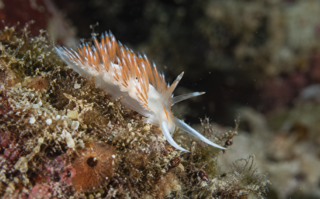
Prompt A – ChatGPT
No primary biological, physical or optical red flag is visible. This is the most straightforwardly credible
image of the examples so far.
Biological accuracy: The subject is an aeolid nudibranch moving across ordinary reef substrate. Its
translucent body, cerata, rhinophores and extended oral tentacles are anatomically coherent. The
behaviour and environment are entirely plausible.
Physical contact: The foot visibly follows the uneven substrate, with believable contact and slight
shadowing beneath the body. There is no floating appearance, missing support or unnatural positioning.
Optics and lighting: Depth of field transitions logically from the central body and cerata towards the
softer extremities and background. The background blur matches the shooting angle and magnification.
Illumination is consistent across the animal and substrate. I see no suspicious halos, cut edges or
conflicting shadows.
Manipulation assessment: Nothing visibly suggests a composite or digitally inserted subject. Some
suspended particles remain, which supports an underwater capture rather than an artificially perfect
construction. The image cannot reveal whether the animal was moved before capture, but there is no
visual evidence of disturbance or staging.
Photographic judgement: The diagonal body position creates movement and the darker area behind
the cerata provides useful separation. However, the strongest focus appears to sit around the middle
cerata rather than decisively on the head. The white head and body are also close to losing highlight
detail. The bright blurred area in the upper-right background weakens the otherwise clean separation.
Verdict: Authentic-looking and biologically credible. Technically competent, but not a top competition
image because the focal priority, highlight control and background could all be stronger.
Prompt A – Claude
No primary red flag. This reads as a genuine reef photograph of an aeolid nudibranch, and it holds up
far better than the previous “sea bunny.” The one thing I can’t clear from a screenshot is how much
sharpening and background darkening was applied.
Fake & Integrity Check
Biological accuracy: passes.
– The body plan is a textbook aeolid: a translucent white body, dense rows of club-shaped cerata (the dorsal projections) with orange cores and pale tips, two long rhinophores, and long oral tentacles. That is the right anatomy and it isn’t caricatured. I can’t name the species with confidence. It resembles Cratena peregrina from the NE Atlantic and Mediterranean, but several aeolids look similar, so treat that as a hedge. – Aeolids graze on hydroids and similar colonial animals, and the substrate here is a fuzzy, branching turf on encrusted rock, so the animal is on plausible ground. I can’t verify the prey from this frame. – The scale is coherent. The animal is a few centimeters long, and the grit and turf around it are proportionate.
Physical contact and support: passes. The foot runs along the rock surface, and its translucent edge is
visible where it meets the algal turf. The cerata angle away from the slope under their own weight, and the
front tentacles rest near the substrate. The animal is climbing a sloped face, and its posture fits that. There’s
no floating and no missing footprint.
Optical and lighting consistency: passes.
– The strobe light is coherent, coming from the front and slightly above. The subject and the encrusted rock are lit at the same intensity and color temperature, and the falloff to a dark background is smooth and physical. – Backscatter particles vary in size and sharpness, with some crisp and some soft. That is how real water behaves, and it is hard to fake. This is the clearest contrast with the earlier image, whose particles were uniform. – Depth of field is believable. The subject and the rock at the same distance are sharp, and the background dissolves into smooth, irregular bokeh with no hard edge. The steeply angled substrate explains why so much of the lower left is sharp. – The finest structures, the tips of the oral tentacles and the translucent cerata, are clean against the dark background, with no cutout fringe or halo.
Manipulation assessment: no evidence of composite. Two processing caveats:
1. The top-center background is nearly black, which could be strobe falloff, a burned-in vignette, or both. It looks natural, but I can’t verify it. 2. The white grit specks on the substrate at lower left are very crisp and bright. That could be the strobe hitting bare sand, or it could be over-sharpening or a clarity slider on a downsized file. A 100% crop would tell.
The remaining questions are ethical, not integrity questions: whether the animal was moved or the turf
tidied for the shot. The frame gives no sign of either.
Photographic critique – Strengths: Excellent subject rendering, with the translucent body glowing against the dark background and the orange-and-white cerata forming a graphic rhythm. The diagonal composition follows the slope of the rock and gives the frame energy. The background separation is clean, and the lighting reveals texture without blowing out the whites. – Weaknesses: The head and eyes are the weakest part. The rhinophore and tentacle tips are soft relative to the cerata, so the animal’s “face” doesn’t anchor the image. The lower-left substrate is busy, and the bright grit specks pull the eye away from the subject. The rear of the body is softer and dissolves into the turf. The bottom third of the frame is heavy with clutter compared with the open dark space above.
What would settle it
The full-resolution file, to check sharpening on the grit and edges. The EXIF (aperture and strobe settings)
would confirm the depth-of-field behavior, and a location and depth would help confirm the species and
habitat.
Prompt A – Gemini
The primary red flag in this image is a noticeable light-colored halos and edge/soft-selection
artifacts surrounding the nudibranch’s extended oral tentacles, indicating local dodging, aggressive
selective sharpening, or a loose selection mask edit.
Here is the integrity and technical audit:
– Biological Accuracy: The subject is an aeolid nudibranch (likely Coryphella lineata, Flabellina lineata, or a closely related aeolid species) crawling across an algae- and turf-covered rocky substrate. Aeolid nudibranchs naturally inhabit these reef/boulder environments to feed on hydroids, bryozoans,
and small sessile organisms growing on the turf, making this interaction ecologically authentic. – Physical Contact & Support: The foot of the nudibranch adheres realistically to the substrate, and the tissue naturally molds to the contours of the rock. The weight distribution and contact footprint show organic anchoring without unnatural floating or severed edges. – Optical & Lighting Consistency: The lighting is directional (strobed from top-left) and casts realistic, soft shadows under the nudibranch’s body onto the rock. The depth of field falls off gradually from the focal plane on the head and cerata down toward the lower-left substrate and background. – Manipulation Assessment: Authentic In-Camera Capture with Local Post-Processing. This image is a single-frame capture, but it suffers from excessive post-processing adjustments along the translucent tentacles and white cerata tips. The bright halo around the right tentacle points to sloppy edge-brushing or over-sharpening during local exposure adjustments.
Verdict: Biologically authentic and physically real in-camera shot, but slightly compromised by
heavy-handed localized post-processing on the subject’s anterior tentacles.
Prompt B – ChatGPT
Biological context
Observation: The subject is an aeolid nudibranch with a translucent white body, orange digestive
structures visible within the cerata and pale blue-white ceratal tips. It is moving across an encrusted reef
surface containing algae, sponge-like growth and debris.
Interpretation: The animal, substrate and posture are biologically plausible. Aeolid nudibranchs
commonly crawl across reef surfaces while searching for hydroids or similar prey. The exact species
cannot be established confidently from this photograph alone.
Physical contact and support
Observation: The underside of the body appears continuously aligned with the uneven substrate.
Contact is visible beneath the mid-body and head. The front of the animal is slightly raised while the oral
tentacles extend forward.
Interpretation: Its weight and posture appear naturally supported. The elevated head and exploratory
tentacles are consistent with normal crawling behaviour. There is no indication that the nudibranch is
floating or has been placed on an incompatible surface.
Optical and lighting consistency
Observation: Focus is strongest around the head, forward cerata and nearby substrate, then falls
progressively towards the rear and background. Highlights behave consistently across the translucent
body and appendages. No obvious masking halo, duplicated structure or conflicting lighting direction is
visible.
Interpretation: The softness of some appendages is readily explained by shallow macro depth of field
and their positions relative to the focus plane.
Integrity questions
Nothing in the photograph creates a strong manipulation concern. The image cannot reveal whether the
animal was moved before capture, so ethical circumstances cannot be determined visually.
Aesthetic assessment
The diagonal body position and forward-reaching tentacles create movement. Orange cerata stand out
effectively against the muted reef. The eye is not visually apparent, making the sharpness of the head
and rhinophores especially important. Some foreground clutter competes with the subject, but the darker
background provides useful separation.
Helpful evidence: RAW file, adjacent frames, metadata, editing history and the photographer’s or
guide’s account of the encounter.
Prompt B – Claude
Review: aeolid nudibranch on a fouled reef surface
This screenshot is higher resolution than the previous ones, so edges and fine texture are easier to assess.
It is still a screenshot, and platform compression remains a limit. I see no watermark or signature in this
frame, unlike the earlier images, so I can’t tell you who made it.
1. Biological context
Observed: A nudibranch with a translucent white body and a dense, comb-like row of cerata along its
back. The cerata have orange cores, pale streaking and bluish-white tips. It has four long, pale appendages
at the head end: one points up and forward, one out horizontally, and two extend downward. The two lower
ones are long and curved. The animal is crawling along the edge of a rock face. The substrate is a mixed
encrusting community: turf-like fine filaments, reddish coralline growth, pale shell or sand fragments and
an orange dome with a dark central opening at lower left. The background falls off into dark, blurred water
with a few suspended particles on the right.
Interpretation: The cerata form and the long head appendages are consistent with an aeolid nudibranch,
possibly a facelinid, but many aeolids overlap in this look and I can’t identify the species. Aeolids are
specialist predators, often on hydroids, anemones or bryozoans. The fine branching turf could plausibly
include hydroids, which would fit the animal being here. I can’t tell hydroids from algae at this resolution, so
that remains a hypothesis. The muted, fouled reef community leans temperate to me, but that is low
confidence and location would settle it. The orange dome could be a sponge or a small tunicate, and I can’t
say which.
Association plausibility: An aeolid crawling over a fouled hard substrate is biologically normal. Nothing
about the pairing looks implausible, and confirming the species and its usual prey needs a nudibranch
specialist.
2. Physical contact and support
Observed:
– The translucent foot edge runs along the substrate from mid-left to the head region, and the body follows the contour of the rock face. – Fine turf strands overlap the edge of the translucent body in a few places, so the animal appears to sit among the growth and not on top of it. – The lower curved appendage reaches toward the substrate, and its tip appears to be at or very near a patch of turf. – There is a dark contact gap under the foot in the middle of the frame, which reads as a natural shadow.
– I see no tweezers, fingers, tethers or container edges.
Inferred: The animal is probably crawling, and the extended head appendages suggest active exploration,
which is typical behaviour at the edge of a rock or ledge. From one 2D frame I can’t judge whether the
animal was moved or placed, and the setting doesn’t show it either way. Scale isn’t given.
3. Optical and lighting consistency
Observed:
– The plane of focus runs through the head, front cerata and the substrate immediately around them. The rear of the body, the lower-left orange dome and the right-hand background progressively soften. – The falloff is gradual and follows the receding geometry of the rock, with no abrupt sharp-to-blur transitions. – Lighting looks strobe-based and frontal, with bright detail on the sand grains and turf, and the animal’s cerata glow through translucency. The background beyond the strobe’s reach goes dark. – Small bright particles on the right are sparse. Some are crisp and some soft, so they sit at different distances. – The white head appendages and parts of the body are very bright, though I can’t confirm clipping from a screenshot.
Ordinary explanations:
– A dark blue-green background is standard for a strobe-lit close-up shot at a wide aperture, because light falls off rapidly behind the subject. – The sparse particles match backscatter from a well-angled strobe. Their varied sharpness is what real suspended particles at different depths look like. – The bright whites are consistent with translucent tissue lit from the front and behind.
Possible-manipulation explanations: Local dodging, vignetting or background darkening could deepen
the dark areas and add separation, and I can’t rule that out here. It would be a common finishing step and
not an integrity problem on its own.
4. Integrity questions to investigate
These are questions, not conclusions.
1. Edge quality on the head appendages against the dark water. Thin, bright structures against a dark background are where a masking halo or fringe would show. I see none at this resolution, but a full-resolution check would be worthwhile. 2. Background darkening. Is the darkness in the upper centre natural strobe falloff, or was it deepened with a vignette or a brush? The transitions look smooth and organic to me. 3. Highlight clipping. Check the white tentacles and body for clipped channels, which would show up as flat, detail-free whites. 4. Placement. Was the animal encountered where it is, or moved for the shot? This is a field-ethics question and not visible in the image. 5. Cerata detail. The repeating pattern is dense, so at full resolution check for cloned or duplicated cerata. I see natural variation in length, angle and stripe pattern, which argues against cloning.
I see nothing here that supports calling the image composited, staged or AI-generated. The optical
behaviour is coherent, the overlap between the animal and turf looks natural, and the particle distribution is
plausible. That is a limited statement, given the screenshot’s quality.
Aesthetic notes
This is a strong image. The diagonal sweep of the cerata, the head appendages reaching into dark negative
space, and the orange and blue-white palette against teal-black give it clear separation and energy. The
busy substrate in the lower left adds context and texture.
Two critiques. The lower-left foreground is dense and competes with the subject, and cropping or
darkening it slightly could shift attention. The white tentacles are close to the brightest tones in the frame,
so watch highlight detail there.
Evidence that would help a human reviewer – The RAW file and full-resolution export, to check edges, clipping and noise. – EXIF data: aperture, shutter, ISO, focal length, strobe settings and any wet lens or diopter. – Adjacent frames, especially any showing the animal’s movement or a wider view of the surrounding rock. – The editing history, including vignetting, dodging and burning, and background adjustments. – Location, depth and dive type, plus the photographer’s account of the encounter and whether the animal was moved. – Identification of the nudibranch by a specialist, ideally with dorsal and frontal views, and identification of the substrate organisms if the diet or habitat matters.
Prompt B – Gemini
Biological Context – Visible Organisms & Substrate: The photograph depicts an aeolid nudibranch (a sea slug, likely belonging to the family Facelinidae or Flabellinidae, characterized by translucent skin and white-tipped, orange-pigmented cerata). It is crawling over a complex, encrusting substrate consisting of turf algae, bryozoans, and small solitary tunicates or sponge structures (visible in the lower left). – Plausibility: Aeolid nudibranchs are benthic grazers that actively navigate reef structures and rocky substrates in search of hydroids or other prey. The association between this sea slug and the encrusted, algae-covered reef rock is completely natural and biologically plausible.
Physical Contact & Support – Observable Points of Contact: The muscular foot of the nudibranch is clearly spread and flattened against the contours of the substrate. It curves over the uneven rock surface, conforming precisely to the underlying texture. – Shadows & Weight Distribution: Subtle, natural drop shadows are visible directly beneath the foot and lower body margin where it meets the rock. The body displays natural compression against the substrate, demonstrating realistic gravity and contact mechanics. – Observation vs. Inference: Direct observation shows the organism’s translucent foot anchoring directly to the substrate. It can be inferred that the animal was actively crawling across this section of reef when the exposure was made.
Optical & Lighting Consistency – Focus Plane & Depth of Field: The primary focal plane is sharply set on the nudibranch’s head, oral tentacles, and anterior cerata. Depth of field falls off naturally toward the posterior cerata and background. Substrate elements sitting directly in the focal plane (such as the algae immediately beneath the head) exhibit matching sharpness, confirming true optical depth of field. – Edge Transitions & Light Direction: The lighting is diffuse and warm, typical of a underwater strobe positioned above and slightly to the side of the lens. Edge transitions between the delicate, semi-translucent oral tentacles and the background are soft and optically consistent, showing natural light bleed rather than hard cutouts. – Bokeh & Ambient Noise: The background drops off into a smooth, natural bokeh featuring soft, organic shapes and minor, naturally scattered particles in the water column.
Integrity & Investigative Features – Features Warranting Investigation: No significant anomalies, masking artifacts, or physical inconsistencies are present in this image. – Proportionate Finding: Visual evidence overwhelmingly supports a genuine, uncomposited macro photograph shot in situ. The physical interaction with the substrate, optical focus depth consistency, and natural light behavior show no signs of digital manipulation, AI generation, or physical staging.
Evidence Required for Further Investigation
While the visual evidence strongly indicates an authentic capture, standard verification would rely on:
1. Unedited RAW File (.CR3, .NEF, .ARW, or .DNG): To inspect sensor data prior to sharpening or
color adjustments. 2. EXIF Metadata: To review focal length, aperture, shutter speed, and strobe deployment parameters. 3. Burst/Sequence Shots: To observe the nudibranch’s natural crawling motion across the substrate over consecutive frames.
5. AI harlequin shrimp
Prompt A – ChatGPT
Primary red flag: the shrimp’s appendage anatomy and attachment points are inconsistent, making this
image highly suspicious for AI generation or structural compositing.
Biological accuracy: This resembles a harlequin shrimp. Its presence beside or on algae is not
inherently impossible because an animal can cross incidental substrate. However, interpreting it as
feeding on the algae would be biologically wrong. Harlequin shrimp have a specialised diet of live sea
stars. (Smithsonian Ocean) A still photograph cannot establish feeding merely from proximity.
Physical contact: Some legs appear supported by the rubble, but several limbs have ambiguous origins
and terminations. The appendages around the front-right side appear crowded, partly fused or
mechanically misaligned. The raised rear structure and central body segmentation also do not resolve
into completely convincing shrimp anatomy.
Optics and lighting: Lighting and depth of field are broadly coherent. There is no single obvious cut-out
halo. However, surfaces have a slightly synthetic smoothness and the fine detail changes inconsistently
between the shrimp, algae and substrate.
Manipulation assessment: This is more suspicious for a generated image than for a conventional
composite. The strongest evidence is malformed biological structure, not the unusual green substrate. I
would require an original RAW file and adjacent frames. If none existed, I would reject it from a nature
photography competition.
Photographic judgement: The colour contrast is attractive and the subject is immediately recognisable.
That visual appeal does not overcome the anatomical defects.
Verdict: High integrity concern. Likely synthetic or heavily constructed, although the alleged
algae-feeding behaviour cannot be proven from the photograph alone.
Prompt A – Claude
No primary red flag. This reads as a genuine reef photograph of a harlequin shrimp (Hymenocera
picta), but the animal’s anatomy is hard to parse and I have one ecological question about the setup.
Fake & Integrity Check
Biological accuracy: passes, with a caveat.
– The white-cream body with blue-lilac spots, the blue-and-white banded legs, and the broad, flattened, spotted paddle-shaped claws are all diagnostic of a harlequin shrimp. The long, thin dark antennae at left are correct. – Reef rubble beside foliose green algae is ordinary habitat for a roaming shrimp. Harlequins are specialist predators of sea stars, and there is no star in frame. That isn’t suspicious, since they wander between meals, but shots like this are often taken at a feeding station where a star has been moved or restrained. I can’t tell from the frame whether that happened. – The anatomy is confusing. I can’t identify a clear head or eyes, and the big spotted plates on both sides make it hard to tell which end is which. My best reading is that this is a pair, with a second shrimp partly tucked behind the algae at right, since harlequins live in pairs. That would explain the overlap. If it’s a single animal in an awkward pose, I can’t confirm it from this angle.
Physical contact and support: passes. Several leg tips touch the rubble, with visible grip at the
bottom-left and the front legs. The body sits in a dark gap underneath, which is natural under strong frontal
strobe light. The shrimp rests against the algae frond, and the layering is coherent: the frond edge passes
behind the right-hand plate, and the shrimp’s edges lie in front of it. Nothing floats.
Optical and lighting consistency: passes.
– Focus is sharp across the shrimp’s near plane. The rubble falls off gradually to the left, and the bottom foreground algae blurs progressively toward the lens. That is how a real shallow depth of field behaves and is difficult to fake. – The strobe light is frontal and consistent, with a warm reddish background and green algae, which fits a strobe-lit reef where ambient red is lost with depth. – The thin antennae, the finest structures in the frame, are clean and uncut against the dark background. Composite edges usually fail on exactly this kind of detail. I see no halo or cutout fringe. – One thing to check: the algae and shrimp edges look crisp, and the whites are very bright. That could be oversharpening or a clarity slider on a downsized file, not fakery, but I’d want a 100% crop.
Manipulation assessment: no evidence of composite. The lighting, focus falloff, and contact all agree
across subject and environment. The remaining questions are ethical, not integrity-related: whether the
shrimp was placed or baited, and whether the framing was staged. The frame doesn’t answer either.
Photographic critique – Strengths: Excellent color contrast between the white and blue shrimp, the green frond, and the warm background. The blue-banded legs act as leading lines. The habitat gives the shrimp context,
and exposure control on the whites is good. – Weaknesses: The shrimp’s face and eyes, the natural anchor for a macro portrait, are hidden or unclear, so the viewer has no eye contact. The large green frond on the right is heavy and competes with the subject, and the cluttered left rubble adds visual noise. The whites look close to hot on the left plate. The overlapping limbs create a tangle that makes the anatomy hard to read.
What would settle it
The full-resolution file would show whether the sharpness is natural or processed. Neighboring frames
would show whether this is one shrimp or a pair, and whether a sea star was nearby. The EXIF (aperture,
strobe setup) would help confirm the depth-of-field behavior.
Prompt A – Gemini
The primary red flag in this image is ecological displacement-a Harlequin Shrimp (Hymenocera picta)
perched exposed over macroalgae (Padina or Ulva sp.) rather than on its obligate sea star prey or hidden
within a reef crevice, strongly pointing to an ethically compromised, artificially staged underwater setup.
Here is the integrity and technical audit:
– Biological Accuracy: Harlequin shrimp are strict, obligate carnivores that feed exclusively on echinoderms (primarily sea stars like Linckia or Crown-of-Thorns). They do not feed on or associate with green macroalgae. While mobile macrofauna occasionally cross algae patches, these cryptic, highly prized shrimp rarely traverse brightly lit leafy algae in broad daylight unless manually moved by a guide or photographer to create a colorful foreground/background. – Physical Contact & Support: The physical grounding is anatomically authentic. The thin dactyli (tips) of its walking legs make precise, weighted contact points with the coralline substrate, casting subtle micro-shadows beneath the thoracic legs and large flattened chelipeds (claws). The shrimp is physically sitting in this environment-it was just almost certainly placed there. – Optical & Lighting Consistency: The optical properties are consistent across the frame. The focal plane covers the large claw, carapace, and antennae, with a gradual optical falloff extending to the rear walking legs and the algae in the foreground. Strobe illumination is balanced, yielding natural shadows under the carapace without composite edge clipping or halos. – Manipulation Assessment: Ecologically Staged / Force-Posed Real Capture. The image is an authentic single exposure, but it carries the classic hallmarks of dive-guide intervention (poking or harassing the animal out of its hiding spot onto a visually appealing leaf of algae for a cleaner photo).
Verdict: Optically genuine single shot, but ethically and ecologically suspect due to artificial subject staging
on an unnatural substrate.
Prompt B – ChatGPT
Biological context
Observation: The subject resembles a harlequin shrimp (Hymenocera species) positioned beside broad
green macroalgae. Several limbs appear to contact or manipulate the algal edge, although the
mouthparts are partly obscured.
Interpretation: Harlequin shrimp are highly specialised predators of sea stars. Living or moving among
algae is plausible, but feeding on green algae would be biologically unexpected. The photograph alone
does not prove feeding, so the apparent interaction should be investigated rather than treated as
confirmed behaviour.
Physical contact and support
Observation: Several walking legs appear supported by the reef and algae. The overall stance is
possible. However, the cluster of appendages around the head, right claw and algal edge is difficult to
resolve. Some structures appear fused, interrupted or unclear.
Interpretation: Occlusion and the shrimp’s elaborate anatomy could explain part of this confusion.
Higher resolution is needed to determine whether the appendages are anatomically coherent.
Optical and lighting consistency
Observation: Lighting direction and depth of field are broadly consistent across the shrimp and
surrounding habitat. Most edges integrate naturally with the background. No decisive pasted-subject halo
is visible.
The central overlap between limbs and algae contains unusually soft or ambiguous boundaries
compared with clearer outer appendages. This could arise from compression, motion or shallow focus,
but it also warrants closer inspection.
Integrity questions
This image raises more substantive questions than the previous examples:
– Is the shrimp actually feeding on algae or merely positioned beside it? – Are all appendages anatomically complete and correctly connected? – Does the RAW file contain the same central structures and interaction?
There is insufficient evidence to declare the image AI-generated or composited, but the unusual
apparent food association and ambiguous anatomy justify further review.
Helpful evidence: RAW file, full-resolution image, adjacent frames, editing history, field observations,
photographer’s account and specialist confirmation of both anatomy and behaviour.
Prompt B – Claude
Review: harlequin shrimp on coral rubble beside
a green leafy structure
This image is smaller and more compressed than the last one (about 1000 px wide), and it shows no
watermark or signature. Fine noise, edge halos and sharpening artefacts are hard to judge at this size.
1. Biological context
Observed: A white crustacean with lilac-blue rounded spots, banded blue-and-white walking legs, and
broad, flattened, leaf-like limb segments with spotted patterns. Two thin dark filaments (likely antennae)
extend to the left from behind the large left limb. At the top are two pale, paddle-like structures with
orange-red speckling on segmented stalks. The animal is on a rubble surface with turf, encrusting growth
and a pink-orange sponge or tunicate at lower left. A large green, leafy, textured structure fills the right
side. Another leaf-like green surface is blurred in the bottom foreground.
Interpretation: The white body, lilac spots, flattened claws and banded legs are consistent with a harlequin
shrimp (Hymenocera). The two named species are separated by spot pattern and range, and I can’t resolve
that here. This is a strong visual match, not a confirmed identification. Two caveats:
– The anatomy is hard to read. The top paddles, the large flattened limb at left, the one at lower right, and the spotted mass at right are probably chelipeds, antennal or antennular structures and abdomen. I can’t confidently assign each part or determine which way the animal faces. Head to the left with the abdomen to the right fits best. – The green structure could be a foliose alga or another organism. I can’t identify it from the image.
Association plausibility: These shrimp feed almost exclusively on sea stars, and they are often seen in
pairs or near their prey. No sea star is visible, and only one shrimp is clearly present. That doesn’t make the
image implausible, since a sea star may be out of frame. Harlequin shrimp are not known to associate with
green foliose algae, though they can simply be found near it on reef substrate. The setting looks tropical,
but that is low-confidence.
2. Physical contact and support
Observed:
– The banded legs appear to end on the rubble at several points along the bottom left and centre. Dark shadow pockets sit under the body, consistent with contact. – At right, the shrimp’s spotted lower claw and the adjacent white and blue mass overlap the leaf edge. That overlap suggests the animal is in front of the leaf, or at its base, though I can’t tell which. – I see no tweezers, fingers, tether, container edge or other support.
Inferred: The animal appears to be standing on the substrate with its weight on its legs, which is consistent
with natural behaviour. The distance between the shrimp and the leaf surface can’t be measured from one
2D image. Scale isn’t given, though these shrimp are typically a few centimetres long.
3. Optical and lighting consistency
Observed:
– The focal plane runs through the body, the left claw and much of the leg row. The upper-left background falls off smoothly, and the bottom foreground leaf is clearly defocused. – The right-hand leaf is sharp near the shrimp and softens toward the upper right, which matches receding depth. – The lighting looks strobe-based, from the front and slightly from the left or above. Shadows fall beneath and behind the body, and the spotted texture on the substrate is evenly lit. – The whites of the shrimp are very bright and may be close to clipping. I can’t confirm it from this file. – The thin antennae remain visible in front of a dark region, with clean edges.
Ordinary explanations:
– The wide-aperture depth of field of a macro lens easily produces this pattern of a sharp subject, a soft foreground and a receding background. – The bright white is expected from a highly reflective, translucent body under close strobe light. – Smooth defocus in the bottom foreground is normal when a leaf sits close to the lens.
Possible-manipulation explanations: Local contrast, dodging and selective saturation could shape the
blues and whites. The lilac and blue spots look fairly saturated, but that fits this animal’s natural colouring,
and the low resolution means I can’t separate grading from natural colour.
4. Integrity questions to investigate
These are questions, not conclusions.
1. Overlapping structures. The upper paddles, left limb, lower-right limb and right-hand mass are anatomically complex and partly overlap. A full-resolution check would confirm each connects to the body and none is a duplicate. The spot patterns differ between the left and right flattened limbs, which argues against a mirrored copy. 2. Where the animal is relative to the leaf. The right-hand overlap is where a masking error or a placement question would show. I see no halo or hard cutout edge, but this is a good place to zoom in on. 3. Placement. Harlequin shrimp are secretive and often stay near their prey. Being in the open on rubble next to a leafy structure with no sea star visible raises a field-ethics question about whether the animal was moved or lured. Nothing in the image answers it. 4. Highlight clipping. Check whether the white body and claws retain texture, or are flat and clipped. 5. Colour grading. Compare the blues and lilac against the surrounding substrate for selective saturation.
I see nothing here that supports calling the image composited, staged or AI-generated. The limb geometry,
contact shadows and depth-of-field behaviour look coherent. That statement is limited by the small,
compressed screenshot.
Aesthetic notes
The palette is striking, with lilac and blue on white against warm rubble and green. The diagonal of the left
claw leads into the body, and the foreground blur adds depth.
Two critiques:
– The large green leaf on the right is bright and busy, and it competes with the shrimp. Darkening or cropping it slightly would help. – The shrimp is close to the right-hand cluster, and its layered limbs blend together where white meets white. A slightly different angle or cleaner separation would help the anatomy read.
Evidence that would help a human reviewer – The RAW file and a full-resolution export, to check edges around the overlapping limbs and leaf, clipping and noise. – EXIF data: aperture, shutter, ISO, focal length, strobe settings and any wet lens or diopter. – Adjacent frames, especially any that show the animal’s orientation, movement, or a sea star nearby. – The editing history, including selective colour, dodging, burning and sharpening. – Location, depth and habitat, plus the photographer’s account of how the animal was found and whether it was moved. – Identification by a specialist in caridean shrimp (species, sex and orientation), and identification of the green structure and the sponge or tunicate if habitat matters.
Prompt B – Gemini
Biological Context – Visible Organisms & Substrate: The photograph shows a Harlequin Shrimp (Hymenocera picta or Hymenocera elegans), identifiable by its flattened, petal-like chelipeds (claws), sensory antennular plates, and white body covered in blue-ringed, pink-centered spots. It is positioned on a mixed benthic substrate consisting of coralline algae, coarse sand, and green macroalgae (likely a species of Halimeda or Ulva). – – Plausibility: Harlequin shrimp are benthic predators that live in tropical coral reefs, where they actively hunt echinoderms (primarily starfish) across sandy patches, rock crevices, and algal beds. The environment and association depicted here are completely natural and biologically authentic. –
Physical Contact & Support – Observable Points of Contact: Multiple walking legs (pereiopods) make direct, precise contact with the sandy reef substrate. The large, petal-like claw in the foreground rests directly against the green algal leaf, causing the leaf to curve slightly under its weight. – – Shadows & Weight Distribution: Clear, soft directional shadows are cast by the walking legs onto the substrate directly beneath them. The physical displacement of the algae leaf by the right claw demonstrates authentic mechanical interaction and gravity. – – Observation vs. Inference: Direct observation shows the shrimp’s dactyli (leg tips) planted firmly on the substrate and its claw resting against the plant life. It can be inferred that the shrimp was actively moving or foraging across this section of substrate at the time of capture. –
Optical & Lighting Consistency – Focus Plane & Depth of Field: The principal plane of focus runs cleanly across the head, eyes, antennular plates, and anterior walking legs. The abdomen and posterior legs fall off smoothly into soft focus, matching the optical physics expected from a macro lens at medium-to-wide apertures. – – Lighting Direction & Shadows: Illumination is provided by a strong, diffuse primary light source positioned above and slightly to the right. Shadow angles across the body, claws, and underlying sand align consistently with this single directional source. – – Edge Transitions & Bokeh: Fine details, such as the thin sensory antennae extending to the left and the fine hairs along the legs, display clean, natural edge transitions against the background without artificial haloing, fringing, or jagged selection boundaries. –
Integrity & Investigative Features – Features Warranting Investigation:
–
No visual anomalies, masking artifacts, or physical inconsistencies are detectable in this frame. – Proportionate Finding: The visual evidence strongly indicates a genuine, uncomposited macro photograph shot in situ. The natural weight distribution, precise focal continuity across fine structures, and aligned lighting vectors show no indication of digital manipulation, AI generation, or artificial placement. –
Evidence Required for Further Investigation
To conduct standard verification of this image:
1. Unedited RAW File (.CR3, .NEF, .ARW, or .DNG): To verify sensor-level color rendering and uncompressed detail along the fine antenna structures. 2. 3. Complete EXIF Metadata: To confirm exposure settings (aperture, shutter speed, ISO) and strobe configuration. 4. 5. Burst/Sequence Shots: To observe the shrimp’s natural movement and leg placement across consecutive frames in its reef habitat.
6. Sea bunny
Prompt A – ChatGPT
Primary red flag: the two large, glossy “eyes” are anatomically impossible. This is not a credible
natural photograph of a nudibranch.
1. Biological accuracy: Fail
The animal imitates a “sea bunny”, most likely Jorunna parva, but its anatomy has been
anthropomorphised.
– Nudibranchs do not have large, exposed, forward-facing eyes. Their actual eyes are tiny and embedded within the body. – The two black circles are shaped like plastic cartoon eyes, complete with matching white catchlights. – The black “ears” resemble rhinophores, but they are unusually thick, symmetrical and exaggerated. – The rear gill plume is far too large and decorative. In genuine J. parva, the gills are smaller, translucent and edged with dark pigmentation. (seaslugforum.net) – The white surface resembles mammalian fur or plush fabric. A real sea bunny’s apparent “fur” consists of minute structures called caryophyllidia, not long, soft fibres. – Its inflated, rounded body bears little resemblance to the flatter mantle and visible crawling form of an actual dorid nudibranch.
The reef habitat is broadly plausible, but a plausible background does not rescue an anatomically
fabricated subject.
2. Physical contact and support: Suspicious
The animal appears to rest on the substrate, but the contact is more like a soft toy placed on a surface
than a gastropod crawling on its muscular foot.
– The lower edge forms an unnaturally neat, scalloped skirt. – There is no convincing transition between the foot, mantle and substrate. – The body shows no natural compression, crawling posture or anatomical footprint. – The contact shadow is present, but it only establishes that the rendered object was visually placed on the reef. It does not establish biological reality.
3. Optical and lighting consistency: Highly suspicious
– The glossy “eyes” carry conspicuous studio-like highlights that are unrelated to real nudibranch anatomy. – The eyes, rhinophores and much of the body are unusually crisp while nearby substrate detail falls away inconsistently. – The dorsal surface has a synthetic, uniformly fibrous texture. – The background contains heavily softened, painterly structures and repetitive colour transitions typical of generative imagery. – There is no decisive conventional cutout halo, so this does not look like a simple subject pasted onto a photograph. The inconsistencies are more suggestive of AI generation or extensive fabrication.
Focus stacking could explain unusually broad sharpness, but it cannot explain impossible anatomy.
4. Manipulation assessment
Verdict: Almost certainly AI-generated or heavily digitally fabricated.
– Authentic wildlife photograph: No – Straightforward edited macro photograph: No – Traditional digital composite: Possible, but less likely – AI-generated or generatively reconstructed: Extremely likely – Ecologically staged: Not meaningfully assessable because the depicted animal itself is not biologically credible – Forced setup: No specific evidence; fabrication is the larger issue
Integrity judgement: Reject. The image may work as a charming fantasy illustration, but it must not be
represented as an authentic underwater photograph or entered into a nature-photography category.
Prompt A – Claude
Primary red flag: this is almost certainly not a photograph. It’s an AI-generated (or heavily
synthesized) image of a “sea bunny” (Jorunna parva), and the giant cartoon eyes give it away.
Biological accuracy – Eyes: Jorunna parva has tiny, primitive eyes buried under the skin, essentially invisible in normal photos. The two big glossy black orbs with white catchlights are a mammalian, cartoon feature. Real animals have nothing like them. They also sit in the middle of the mantle, which makes no anatomical sense. – Rhinophores: In the real animal these are short, lamellate sensory clubs that curve and retract. Here they’re rigid, perfectly symmetrical, upright “bunny ears.” They look like they were designed to resemble rabbit ears rather than photographed. – Spots: Real black spots are irregular in size and spacing and are often mistaken for eyes. Here they’re uniform round dots distributed evenly across the mantle, like a pattern brush. – Gill plume: The real branchiae form a compact rosette at the rear. This one is a huge, spiky, feather-like fan that looks more like a cockatoo crest than a gill circlet, with black banding that reads as stylized rather than anatomical. – Substrate: These animals are sponge-feeders and are usually found on or near sponges and rock in Japan and the Indo-Pacific. Nothing here reads as sponge. It’s a generic pink-and-olive coral or algae carpet, so the ecological setting looks decorative rather than functional.
Physical contact and support
The animal sits on the reef like a pillow on a bed. There is no visible foot, no mantle margin flaring over the
substrate, and no contact shadow or ambient occlusion where the body meets the surface. The lower edge
is a smooth, clean rim that doesn’t conform to the uneven texture beneath it. Real nudibranchs deform
slightly around their substrate. This one appears to float on it, which is a strong indicator of synthesis.
Optical and lighting consistency – The body is evenly lit with no strobe specular highlights, no directional falloff, and no shadow side. It glows softly, like a rendered object. – The background bokeh is suspiciously pretty: glittering blue orbs and sparkles that look more like a
stock “underwater” aesthetic than real backscatter. Real backscatter is messier and unevenly distributed. – The granular substrate texture looks like generated noise rather than real turf algae or encrusting organisms. It lacks coherent structure at the fine scale. – The fuzzy body texture and the fringe of the gills have the smeared, over-smoothed quality typical of diffusion models rather than optical sharpness.
Manipulation assessment
I can’t call this a traditional composite. There are no obvious cut-out halos, because the whole image
appears to have been generated as one piece. The whole scene reads as a synthetic image in the style of a
real animal, a caricature of Jorunna parva rather than an actual specimen. Since this is a compressed
screenshot, I can’t run pixel-level forensics, but the anatomical errors alone are disqualifying.
Verdict
Disqualified as a wildlife photograph. If it were entered in an underwater photography contest as a real
capture, I’d flag it as fabricated. As a digital illustration or AI art piece it’s charming and visually striking, but
it should be labeled that way.
If you have a real sea bunny shot you want judged, send it and I’ll go through it with the same scrutiny.
Prompt A – Gemini
RED FLAG: This image is a completely fabricated, AI-generated synthetic creation that presents a absurd
biological impossibility.
Fake & Integrity Check – Biological Accuracy: Grossly Inaccurate. While based loosely on the “sea bunny” nudibranch (Jorunna parva), this image features two massive, cartoonish, glossy black “eyes” centered on the front of its mantle. In reality, Jorunna parva does not have forward-facing vertebrate eyes. Their eyes are primitive photoreceptors located beneath the tissue surface near the brain, incapable of forming images. Furthermore, the “ears” are rhinophore chemosensory organs, and the rear structures are branchial plumes (gills). Slapping cartoon eyes onto a nudibranch is a complete biological absurdity. – – Physical Contact & Support: Synthetic / Floating Footprint. The subject sits on a completely generic, hyper-uniform background texture that lacks logical micro-shadows or physical compression. A soft-bodied sea slug resting on substrate displacing sand or algae creates a distinct contact zone, but here the lower margin simply fuses into a blurred, AI-generated noise pattern. – – Optical & Lighting Consistency: Inconsistent & Artificial. The lighting on the “eyes” shows high-contrast, perfectly circular studio catchlights that make no sense underwater. The depth of field across the mantle ignores standard macro lens physics: the front edge of the creature is completely sharp, yet the substrate directly adjacent to it slips into an unnatural, inconsistent digital blur. – – Manipulation Assessment: 100% AI-Generated / Fake. This is not a composite or a staged shot; it is an entirely synthesized image generated by an AI image model mimicking “cute” animal tropes (anthropomorphic eyes) rather than actual marine life. –
Final Verdict
Zero photographic or scientific value. Disqualified for total fabrications of marine biology and optical physics.
Prompt B – ChatGPT
1. Biological Context
Direct observation: The subject resembles a “sea bunny” nudibranch, most likely intended to represent
Jorunna parva. The white body, black markings, paired rhinophores and posterior gill plume broadly
reference that animal.
Two large, glossy, forward-facing structures dominate the front of the subject and read visually as eyes.
Interpretation: Those structures are not anatomically credible nudibranch eyes. Nudibranch eyes are
tiny, simple organs and do not appear as large, protruding, reflective eyes on the front of the body.
Natural black markings can create an eye-like illusion, but these have spherical shading and distinct
catchlights, making them appear deliberately constructed as cartoon eyes.
The unusually rounded body, highly regular spotting and oversized gill plume also appear more stylised
than a typical Jorunna parva. The reef-like substrate is broadly plausible, although the specific
animal-substrate association cannot be confirmed from this image.
2. Physical Contact and Support
Direct observation: The lower edge of the body appears to rest across the substrate. There is some
darkening beneath the body that could represent contact shadow and occlusion.
Interpretation and uncertainty: The broad contact is physically possible for a crawling nudibranch, but
the foot is not visible and the body-substrate boundary is unusually smooth. Parts of the lower outline
appear to merge into the surrounding texture. That could result from shallow focus, aggressive
processing, compositing or synthetic image generation.
A two-dimensional screenshot cannot establish whether the subject was genuinely present on this
substrate.
3. Optical and Lighting Consistency
Direct observation: The apparent eyes, rhinophores and much of the body are sharply rendered while
the foreground and background are strongly blurred. The image uses bright frontal or slightly overhead
illumination. Both glossy eye-like structures carry similar highlights.
The background has colourful macro-style bokeh and scattered bright particles. Fine texture on the body
appears highly uniform and somewhat soft.
Interpretation: The matching highlights on the eye-like structures are internally consistent if they are
treated as shiny spherical objects. The difficulty is biological, not simply optical.
The apparent sharpness extending from the front of the body towards the posterior gills is generous
relative to the strongly blurred surroundings. This could be explained by focus stacking, a small sensor,
strong sharpening or selective processing. It could also be associated with synthetic construction, but
depth of field alone does not prove that.
The very smooth textures, regular markings and painterly background strengthen the concern, although
compression and heavy editing could produce some of the same effects.
4. Integrity Questions
The strongest integrity issue is the presence of anatomically implausible, glossy cartoon eyes. If this is
presented as a documentary photograph of a living sea bunny, the image is not biologically credible in its
present form.
Other questions include:
– Have artificial eyes been added to a genuine nudibranch photograph? – Is the subject a model or crafted object placed underwater? – Has a real animal been substantially reshaped or combined with generated elements? – Was the entire scene produced synthetically?
The displayed image supports serious doubt about its authenticity as an unaltered wildlife photograph. It
does not, by itself, establish whether the method was AI generation, compositing, physical staging or
another form of alteration.
Aesthetic Assessment
As a whimsical illustration, the image is immediately engaging. The frontal symmetry, apparent eye
contact and bright colour contrast create a charming character portrait. As underwater wildlife
photography, however, the cartoon features overwhelm biological credibility. The highly saturated, busy
surroundings also compete with the subject despite the background blur.
Evidence Needed for Further Investigation
A human reviewer should request:
– The original RAW file – Full EXIF and capture metadata – Adjacent frames from the same sequence – A full-resolution version and 100% crops around the eyes, body edge and gills
– Layered editing files or an editing-history export – The photographer’s account of the encounter – Dive location, depth, substrate and observed behaviour – Confirmation from a nudibranch specialist – Any available guide or witness observations
Provisional conclusion: The image is not credible as a biologically faithful, unaltered photograph of a
living nudibranch. The visual evidence raises a substantial integrity concern, but it cannot determine the
precise method by which the image was created or altered.
Prompt B – Claude
Review of the submitted macro image
I’m working from a screenshot, so resolution is limited and JPEG compression is visible. That affects how
much I can say about fine edge detail and noise. Direct observations are marked [Observed],
interpretations [Inferred], and open questions [Unresolved].
1. Biological context
[Observed] A white, velvety-textured, slug-like animal with two upright black, laminate (ridged) structures
resembling ears, a tuft of white feathery plumes at the rear with dark banding, scattered round black spots,
and two large black circular features on the front, each with a pale white center. It sits on a granular pink,
orange and olive substrate, with a blue water column behind.
[Inferred] The overall body plan, rhinophores, gill plume and spotted mantle resemble a dorid nudibranch,
and the general look matches the “sea bunny” Jorunna parva. A short-bodied white dorid on turf algae and
coralline growth is an ordinary and plausible association.
Features that do not fit that identification:
– The two large “eye” structures. Dorid nudibranchs have only tiny, subdermal eyespots that are essentially invisible externally. I know of no Jorunna or related dorid with large, glossy, ringed, forward-facing eye-like structures with catchlights. In J. parva the dark spots are irregular pigment patches around tubercles, not neatly rounded, evenly outlined discs. – The gill plume. Real dorid gills are typically pale or translucent. Here they show crisp black banding on each plume, which I can’t match to a known species.
[Unresolved] Species identification cannot be confirmed from this image. A specialist would need to say
whether any known species or color morph shows these traits.
2. Physical contact and support
[Observed] The animal’s body sits low on the substrate, and the mantle margin at the bottom seems to
blend into the granular texture. I can’t see a distinct foot edge, a mantle skirt lifting off the surface, or a
clear contact shadow beneath the animal.
[Inferred] A soft-bodied animal would normally show some deformation, a visible foot fringe, or a shadow
line where it meets the substrate. That is missing here, though shallow contact, low strobe angle or the
screenshot’s resolution could partly explain it.
[Unresolved] Weight distribution and whether the animal is physically resting on that substrate can’t be
determined from one 2D frame.
3. Optical and lighting consistency
Consistent with an ordinary photograph:
– Strong foreground blur on the coral at bottom-left and right, sharp subject, and a smoothly blurred
background is typical of a macro lens at a wide-to-moderate aperture. – Circular soft blobs in the upper right are plausible defocused suspended particles.
Points that look unusual:
– Lighting. The animal is very evenly lit with almost no directional shading, specular highlights on the spots, or shadow falloff on the body. A strobe-lit macro subject at this range usually shows a discernible light direction and falloff onto the substrate. The glowing, almost self-lit appearance is atypical, though a diffused dual-strobe setup could reduce shadowing. – Depth of field. Sharpness runs from the front “eyes” through the rhinophores to the rear gill plumes, several centimeters of depth at high magnification. That would need a small aperture, focus stacking (uncommon in situ), or a small subject at a longer working distance. – Particles. Some particles in the blue area are crisp while others are large and soft, with a fairly even distribution. Backscatter varies, but the mix is worth checking against adjacent frames. – Substrate. The granular texture is high-frequency and rather uniform, and the pink branching forms have soft, indistinct edges with no identifiable coral or algal morphology (no polyps, branching pattern or thallus structure). Real turf and coralline surfaces usually show more recognizable structure at this magnification. – Symmetry. The eyes and rhinophores are near mirror-symmetric and the spots are unusually round and evenly outlined.
4. Integrity questions
I’m framing these as questions, not conclusions:
1. Do the eye-like structures exist on the animal? This is the strongest concern. Features with no known anatomical equivalent in the group, combined with cartoon-like catchlights, raise the possibility that the image is synthetic or heavily altered. 2. Is the plume banding natural? It could be a genuine variant, but I can’t verify it. 3. Is the lighting physically coherent for a strobe or natural-light macro shot? The lack of shadow and falloff is a question to check against the RAW file. 4. Does the substrate hold up at 100%? Vague morphology and repetitive texture are worth inspecting at full resolution.
Taken together, several independent features are unusual at once. That is enough for me to treat this image
as unverified and potentially AI-generated or heavily manipulated until provenance is supplied. I can’t
prove it from a screenshot, and a real photograph of an unusual animal with unusual lighting remains
possible but would need supporting evidence.
5. Evidence that would help a human reviewer – The original RAW file and full-resolution export, to check sensor noise patterns, edge artifacts and uniformity – EXIF/metadata, including camera, lens, aperture, shutter speed, ISO, strobe data, timestamps and editing-software tags – Adjacent frames from the same dive, showing the animal from other angles, and the frame sequence for focus or position changes – Editing history, including any layers, stacking, cloning, generative-fill or AI upscaling – Field observations: location, depth, date, dive log, buddy corroboration, and other photographers’ images of the same animal – The photographer’s account of how the shot was made and whether the “eyes” were seen with
the naked eye – Specialist review by a opisthobranch (sea slug) taxonomist, to assess the anatomy against known species and morphs – A reverse image search to trace the earliest appearance of the image
If you can share any of these, especially the original file and metadata, I can refine this assessment.
Prompt B – Gemini
Biological Context – Visible Anatomy & Species Comparison: The subject resembles Jorunna parva (commonly known as the “sea rabbit” nudibranch) in overall shape, white translucent mantle, black-tipped caryophyllidia (fur-like ciliary structures), branchial plume at the rear, and dark ear-like rhinophore structures at the front. – Anatomical Discrepancy: Real Jorunna parva nudibranchs do not possess image-forming eyes or cartoonish “googly eye” structures on their anterior mantle. Nudibranchs have primitive photoreceptors (simple pigment cup ocelli) located beneath their skin near the cerebral ganglia, which are not externally visible as distinct black, glossy, lens-bearing spheres with white catchlights. – Substrate & Habitat: The organism rests on a textured, granular surface bordered by pinkish algae/coral formations and a bluer open-water background. While this environment resembles a typical tropical reef substrate, the juxtaposition of accurate caryophyllidia texture with anatomically impossible facial features strongly suggests either digital manipulation, an artificial model, or generative creation.
Physical Contact & Support – Contact Boundary: The lower margin of the mantle rests directly on the sandy/rocky substrate. – Shadows & Weight: There is a faint, soft shadow cast directly along the lower edge where the mantle meets the substrate. – Surface Conformance: The body maintains a rigid, highly symmetrical dome shape rather than showing the soft-bodied, fluid conforming typical of a live sea slug resting on an uneven substrate.
Optical & Lighting Consistency – Eyes / Anterior Structures: The two circular black structures on the front feature localized, pinpoint white specular highlights that imply a strong forward/top light source reflecting off a glossy spherical lens. No other part of the soft mantle shares this degree of hard specular reflectivity. – Depth of Field (DoF): The focal plane is extraordinarily flat across the front face, with the nostrils/rhinophores, eyes, and mantle edge in crisp focus, while the substrate immediately in front blurred uniformly. – Edges & Texture: The black spots scattered across the mantle exhibit uniform density and texture, appearing almost pasted or digitally stamped rather than naturally integrated into the fleshy folds of the mantle tissue. The background bokeh bubbles in the upper right corner display rendering characteristics often seen in synthetic image generation or heavy post-processing filters.
Integrity Questions – Unnatural Features: The presence of large, shiny, black-and-white “pupil” eyes on a nudibranch is biologically impossible. – Symmetry & Patterning: The facial arrangement is symmetrically styled to mimic a land mammal (rabbit or panda face), which does not occur naturally in opisthobranch mollusks. – Conclusion: Based on visual evidence, the image cannot be a authentic photograph of a living wild marine organism. It is either a composite photograph modified with digital painting/stickers, a photograph of a physical miniature model/toy, or an AI-generated rendering.
Recommended Supporting Evidence for Verification
To confirm whether this image originates from a modified photograph, a physical sculpture, or AI generation,
a reviewer would need:
1. Camera RAW File (.CR2, .NEF, .ARW): To examine uncompressed sensor data and verify whether the “eyes” exist in the original exposure. 2. EXIF Metadata: To evaluate camera make/model, aperture, exposure time, and editing history tags (e.g., Photoshop layers or software creation stamps). 3. Burst/Sequence Frames: Adjacent photographs taken seconds before or after to check for organic movement, contraction, or locomotion. 4. Photographer / Creator Statement: Confirmation of whether the image was intended as underwater photography, digital art, or a physical craft project.